Respiratory Tract DSL: Histology, Structure, Pathology
Basic anatomy and physiology
The respiratory tract has two functional components:
The conducting system, which transports inspired and expired gases between the
atmosphere and the circulatory system.
The interface where passive exchange of gases occurs.
The conducting system begins as a single ‘tube’ which divides repeatedly into
airways of ever decreasing diameter, eventually reaching the blind-end sacs known
as alveoli which are the sites of gaseous exchange.
The respiratory tract is divided into the—
Upper respiratory tract:
-Nose
-Paranasal sinuses
-Nasopharynx
The upper respiratory tract filters, humidifies and adjusts the temperature of
inspired air.
Lower respiratory tract:
-Larynx
-Trachea
-Bronchi/bronchioles
-Alveoli
The lower respiratory tract conducts the air to the sites of gaseous exchange. Vocal
cords are present in the larynx, these prevent large foreign bodies from lodging
within smaller-calibre tubes.
Histology – General Principles
The entire tract is lined by respiratory mucosa, which in turn is composed of
epithelium (‘respiratory epithelium’) and supporting connective tissue (‘lamina
propria). Lymphoid aggregates, which are part of the immune system are present in
the lamina propria.
Deep to the mucosa there is smooth muscle as well as further connective tissue
containing serous and mucous glands and cartilage (around major airways only).
, Nasal mucosa,
low power
There is a gradual transition from one kind of respiratory epithelium to another as
we proceed down the respiratory tract.
In the upper respiratory tract, the larynx and trachea are lined by tall,
pseudostratified columnar ciliated epithelium with many goblet cells.
Cilia are small finger-like processes on the luminla surfaces of the cells, they move
synchronously to waft particles entrapped in mucus towards the pharynx. The goblet
cells produce mucus.
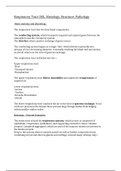
Nasal mucosa,
high power
Basic anatomy and physiology
The respiratory tract has two functional components:
The conducting system, which transports inspired and expired gases between the
atmosphere and the circulatory system.
The interface where passive exchange of gases occurs.
The conducting system begins as a single ‘tube’ which divides repeatedly into
airways of ever decreasing diameter, eventually reaching the blind-end sacs known
as alveoli which are the sites of gaseous exchange.
The respiratory tract is divided into the—
Upper respiratory tract:
-Nose
-Paranasal sinuses
-Nasopharynx
The upper respiratory tract filters, humidifies and adjusts the temperature of
inspired air.
Lower respiratory tract:
-Larynx
-Trachea
-Bronchi/bronchioles
-Alveoli
The lower respiratory tract conducts the air to the sites of gaseous exchange. Vocal
cords are present in the larynx, these prevent large foreign bodies from lodging
within smaller-calibre tubes.
Histology – General Principles
The entire tract is lined by respiratory mucosa, which in turn is composed of
epithelium (‘respiratory epithelium’) and supporting connective tissue (‘lamina
propria). Lymphoid aggregates, which are part of the immune system are present in
the lamina propria.
Deep to the mucosa there is smooth muscle as well as further connective tissue
containing serous and mucous glands and cartilage (around major airways only).
, Nasal mucosa,
low power
There is a gradual transition from one kind of respiratory epithelium to another as
we proceed down the respiratory tract.
In the upper respiratory tract, the larynx and trachea are lined by tall,
pseudostratified columnar ciliated epithelium with many goblet cells.
Cilia are small finger-like processes on the luminla surfaces of the cells, they move
synchronously to waft particles entrapped in mucus towards the pharynx. The goblet
cells produce mucus.
Nasal mucosa,
high power