Neural Control of Breathing
Key Points:
What muscles are used during different types of breathing and how are they
innervated?
How do we breathe without thinking about it? What are we doing when we hold our
breath?
How does the body detect the need to alter ventilation?
What is the difference between central and obstructive sleep apnoea? How do we tell
them apart?
[Involves Lisa case]
In quiet breathing, when we breathe in we use our diaphragm which contracts
(and possibly external intercostals) and when we breathe out this is 100% lung
recoil on its own.
When there is increased ventilation, when we breathe in the diaphragm and
external intercostals contract and when we breathe out there is lung recoil but also
our internal intercostals and abdominals are involved.
Accessory muscles are muscles with other roles that can help with ventilation and
this includes for inhalation, sternocleidomastoid and scalene muscles, for exhaling
our abdominals.
As for the neural innervation of muscles for breathing, it starts in the medulla with
our upper motor neurones. These UMN’s project down to the lower motor
neurones in the spinal cord.
C3-C5 innervates the diaphragm via the phrenic nerve, while T1-T12 innervates
the intercostal muscles.
If there did happen to be a stroke in the area of the medulla where the UMNs
originate then this would completely stop breathing. If there was damage to C3-C5
part of the spinal cord it would cause issues with inhalation due to the diaphragm not
being innervated properly anymore.
Respiratory pattern generator theories (RPG) are theories on how we produce the
breathing that we do. There are multiple theories including: Network theory,
Conditional network bursting theory, pacemaker theory and hybrid pacemaker-
network model.
But at the end of the day, no one actually knows what the real answer is (so no point
learning individual theories). The important thing to remember is just the fact we
have an oscillator for breathing and that it is very complex.
However, everyone does agree that two forms of neurones are important in the RPG,
these are the ventral respiratory group (VRG) which are involved with finishing
inspiration and expiration and the dorsal respiratory group (DRG) which is
involved with inspiration initiation. These originate in the medulla oblongata.
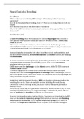
, This is a model of the interaction and functions of these neurones. So it would start
with nucleus 1 (part of the DRG) telling the UMNs to fire to cause the inspiratory
LMNs to fire and thus initiate inspiration. Nucleus 1 will also be signalling to nucleus
3 which then signals all the way up to the VRG and stimulates the UMNs here to cause
the expiratory LMNs to fire and allow exhalation after the inspiration that has just
occurred. There is communication between nucleus 5 + 7 and 7 projects down to tell
nucleus 1 that what it is doing is good and to now inhale again.
There is also inhibition from stretch receptors via the vagus nerve, which tells
nucleus 1 the lungs are full and to stop firing. Note that in normal quiet breathing,
muscles are not required for expiration it is just lung recoil, so to do this the nerves
in the DRG simply have to switch off, only for more heavy breathing are the VRGs
required.
Ventilation can be increased by two different ways, this can be explained by
looking at the equation for VE (V should have a dot above it, indicating flow)
which is the flow you expire (how much you’re breathing basically) –
VE = V T x f
Key Points:
What muscles are used during different types of breathing and how are they
innervated?
How do we breathe without thinking about it? What are we doing when we hold our
breath?
How does the body detect the need to alter ventilation?
What is the difference between central and obstructive sleep apnoea? How do we tell
them apart?
[Involves Lisa case]
In quiet breathing, when we breathe in we use our diaphragm which contracts
(and possibly external intercostals) and when we breathe out this is 100% lung
recoil on its own.
When there is increased ventilation, when we breathe in the diaphragm and
external intercostals contract and when we breathe out there is lung recoil but also
our internal intercostals and abdominals are involved.
Accessory muscles are muscles with other roles that can help with ventilation and
this includes for inhalation, sternocleidomastoid and scalene muscles, for exhaling
our abdominals.
As for the neural innervation of muscles for breathing, it starts in the medulla with
our upper motor neurones. These UMN’s project down to the lower motor
neurones in the spinal cord.
C3-C5 innervates the diaphragm via the phrenic nerve, while T1-T12 innervates
the intercostal muscles.
If there did happen to be a stroke in the area of the medulla where the UMNs
originate then this would completely stop breathing. If there was damage to C3-C5
part of the spinal cord it would cause issues with inhalation due to the diaphragm not
being innervated properly anymore.
Respiratory pattern generator theories (RPG) are theories on how we produce the
breathing that we do. There are multiple theories including: Network theory,
Conditional network bursting theory, pacemaker theory and hybrid pacemaker-
network model.
But at the end of the day, no one actually knows what the real answer is (so no point
learning individual theories). The important thing to remember is just the fact we
have an oscillator for breathing and that it is very complex.
However, everyone does agree that two forms of neurones are important in the RPG,
these are the ventral respiratory group (VRG) which are involved with finishing
inspiration and expiration and the dorsal respiratory group (DRG) which is
involved with inspiration initiation. These originate in the medulla oblongata.
, This is a model of the interaction and functions of these neurones. So it would start
with nucleus 1 (part of the DRG) telling the UMNs to fire to cause the inspiratory
LMNs to fire and thus initiate inspiration. Nucleus 1 will also be signalling to nucleus
3 which then signals all the way up to the VRG and stimulates the UMNs here to cause
the expiratory LMNs to fire and allow exhalation after the inspiration that has just
occurred. There is communication between nucleus 5 + 7 and 7 projects down to tell
nucleus 1 that what it is doing is good and to now inhale again.
There is also inhibition from stretch receptors via the vagus nerve, which tells
nucleus 1 the lungs are full and to stop firing. Note that in normal quiet breathing,
muscles are not required for expiration it is just lung recoil, so to do this the nerves
in the DRG simply have to switch off, only for more heavy breathing are the VRGs
required.
Ventilation can be increased by two different ways, this can be explained by
looking at the equation for VE (V should have a dot above it, indicating flow)
which is the flow you expire (how much you’re breathing basically) –
VE = V T x f