Panniculitides
Lobular: fat lobules (center of adipose tissue lobes). Usually accompanied by vasculitis, but
not always. Examples: lupus panniculitis, pancreatic panniculitis (no vasculitis), erythema
induratum (vasculitis).
Septal: septa (fibrous walls between fat lobules). Example: erythema nodosum.
Erythema nodosum
probably is a delayed hypersensitivity reaction to a variety of antigens.
Epidemiology: It is most common in women in their 20-40s. Women are affected 3-6x more
than men.
Causes
Most common: i) children: streptococcus, ii) adults: streptococcus, sarcoidosis
•Infections (Streptococcus, HBV, HIV, TB)
•Medications (oral contraceptives, penicillins, TNFi [rare])
•Malignancy
•Inflammatory diseases
IBD
Sarcoidosis
Behçet / HA20
RP / VEXAS
Sweet’s syndrome
TAK
•Pregnancy
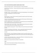
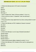
Clinical: tender, warm, erythematous subcutaneous nodules on the bilateral pretibial areas,
young women, 20-40 years. The nodules develop over several days and may follow a
prodromal of fatigue, fever, malaise, arthralgias, or upper respiratory infection symptoms by
1-3 weeks. It has also been reported in other locations (thighs, extensor forearms, rarely,
head, neck, trunk). Ulceration or suppuration of lesions of EN is exceedingly rare. Nodules
last ≈ 2 weeks and then slowly involute without scarring.
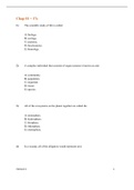
Histology: the characteristic histologic finding in EN is a septal panniculitis without vasculitis.
Septal edema with dominating neutrophils and mild lymphocytic infiltrates. Concomitant
thrombophlebitis may be present, particularly in cases associated with Behçet syndrome.
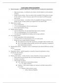
Erythema Induratum (nodular vasculitis, Bazin's disease)
is a lobular panniculitis with vasculitis that frequently occurs in association with TB (and
other infections) and may also occur as an idiopathic condition or in association with other
infections or drug exposure.
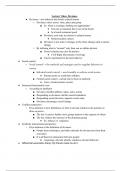
Clinical: it presents clinically as recurrent crops of tender, violaceous nodules and
plaques on the posterior lower legs but lesions have also on the feet, thighs, buttocks and
forearms. The nodules tend to evolve over several weeks, often developing focal ulceration
and drainage. The areas heal with scarring and post-inflammatory hyperpigmentation.
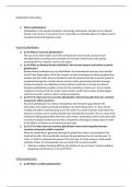
Histology: diffuse septolobular panniculitis with primary neutrophilic vasculitis of
nearby vessels.
Differences: EI has +vasculitis, violaceous and less red than EN, posterior, less painful,
associated more with TB, may ulcerate and may scar.
Lobular: fat lobules (center of adipose tissue lobes). Usually accompanied by vasculitis, but
not always. Examples: lupus panniculitis, pancreatic panniculitis (no vasculitis), erythema
induratum (vasculitis).
Septal: septa (fibrous walls between fat lobules). Example: erythema nodosum.
Erythema nodosum
probably is a delayed hypersensitivity reaction to a variety of antigens.
Epidemiology: It is most common in women in their 20-40s. Women are affected 3-6x more
than men.
Causes
Most common: i) children: streptococcus, ii) adults: streptococcus, sarcoidosis
•Infections (Streptococcus, HBV, HIV, TB)
•Medications (oral contraceptives, penicillins, TNFi [rare])
•Malignancy
•Inflammatory diseases
IBD
Sarcoidosis
Behçet / HA20
RP / VEXAS
Sweet’s syndrome
TAK
•Pregnancy
Clinical: tender, warm, erythematous subcutaneous nodules on the bilateral pretibial areas,
young women, 20-40 years. The nodules develop over several days and may follow a
prodromal of fatigue, fever, malaise, arthralgias, or upper respiratory infection symptoms by
1-3 weeks. It has also been reported in other locations (thighs, extensor forearms, rarely,
head, neck, trunk). Ulceration or suppuration of lesions of EN is exceedingly rare. Nodules
last ≈ 2 weeks and then slowly involute without scarring.
Histology: the characteristic histologic finding in EN is a septal panniculitis without vasculitis.
Septal edema with dominating neutrophils and mild lymphocytic infiltrates. Concomitant
thrombophlebitis may be present, particularly in cases associated with Behçet syndrome.
Erythema Induratum (nodular vasculitis, Bazin's disease)
is a lobular panniculitis with vasculitis that frequently occurs in association with TB (and
other infections) and may also occur as an idiopathic condition or in association with other
infections or drug exposure.
Clinical: it presents clinically as recurrent crops of tender, violaceous nodules and
plaques on the posterior lower legs but lesions have also on the feet, thighs, buttocks and
forearms. The nodules tend to evolve over several weeks, often developing focal ulceration
and drainage. The areas heal with scarring and post-inflammatory hyperpigmentation.
Histology: diffuse septolobular panniculitis with primary neutrophilic vasculitis of
nearby vessels.
Differences: EI has +vasculitis, violaceous and less red than EN, posterior, less painful,
associated more with TB, may ulcerate and may scar.