Unit 2 Biology AS
Topic 3: Cell Structure, Reproduction and Development
3.1 Know that all living organisms are made of cells, sharing some common features
Key Concept:
• The cell theory states that:
o All living organisms are composed of one or more cells.
o The cell is the basic structural and functional unit of life.
o All cells arise from pre-existing cells.
Shared Features of All Cells:
Feature Function
Plasma membrane Controls the movement of substances in and out of the cell
Cytoplasm Site of metabolic reactions
DNA Carries genetic information for controlling the cell
Ribosomes Site of protein synthesis
Enzymes Catalyse reactions essential to life
Energy release All cells must carry out respiration (aerobic or anaerobic) to release energy
Even though structure varies, these core features exist in both prokaryotic and eukaryotic cells.
3.2 Understand how the cells of multicellular organisms are organised into tissues, tissues into organs, and
organs into organ systems
Hierarchical Organisation in Multicellular Organisms:
1. Cells: The basic unit of life (e.g., epithelial cells, muscle cells).
2. Tissues: A group of similar cells working together to perform a specific function.
o Example: Muscle tissue contracts; epithelial tissue lines surfaces.
3. Organs: Made of different tissues working together to carry out a function.
o Example: The heart includes muscle tissue, nerve tissue, and connective tissue.
4. Organ Systems: Groups of organs that work together to carry out a broader function.
o Example: Circulatory system = heart + blood vessels; Digestive system = stomach, intestines,
liver, etc.
,Specialisation:
• Multicellular organisms rely on cell specialisation and cooperation for survival, as no single cell can
carry out all life functions.
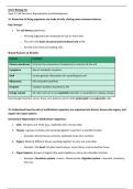
3.3 Ultrastructure of Eukaryotic Cells
(i) Know the Ultrastructure:
Organelle Structure Function
Nucleus Surrounded by nuclear envelope with Stores genetic material; nucleolus
pores; contains DNA and nucleolus makes rRNA and ribosomes
Nucleolus Dense region inside nucleus Synthesises ribosomal RNA
Ribosomes Small, non-membranous; free or bound Site of protein synthesis
to rER
Rough Endoplasmic Membrane-bound sacs with ribosomes Synthesises and processes proteins for
Reticulum (rER) attached secretion or membrane use
Smooth ER Membrane-bound sacs without Lipid synthesis, detoxification
ribosomes
Mitochondria Double membrane, inner folds = cristae, Site of aerobic respiration (ATP
matrix inside production)
Golgi Apparatus Stacks of flattened sacs (cisternae), with Modifies, packages, and transports
vesicles proteins and lipids
, Lysosomes Vesicles containing hydrolytic enzymes Digests old organelles, pathogens, and
waste (intracellular digestion)
Centrioles Cylindrical structures made of Involved in spindle formation during
microtubules (in animal cells) cell division
Note: Only animal and plant cells are eukaryotic. Plant cells also include chloroplasts, a large permanent
vacuole, and a cellulose cell wall.
(ii) Understand Functions of Organelles:
• Protein synthesis and secretion involves coordination of nucleus → rER → Golgi → vesicle → plasma
membrane.
• Organelles provide compartmentalisation, increasing efficiency of metabolic processes.
• Mitochondria generate energy, while lysosomes manage cellular waste.
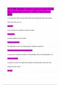
3.4 Role of rER and Golgi in Protein Transport and Enzyme Secretion
Protein Transport Pathway (with modifications and refinement):
1. Transcription in the nucleus – A gene coding for the protein is transcribed into mRNA.
2. mRNA exits the nucleus – The mRNA travels through nuclear pores into the cytoplasm.
3. Translation at ribosomes on the rough endoplasmic reticulum (rER) – Ribosomes on the rER synthesize
the polypeptide chain using the mRNA sequence.
4. Protein enters rER lumen – As the polypeptide is synthesized, it is threaded into the lumen of the rER.
5. Protein folding and modification in the rER:
o The polypeptide folds into its secondary and tertiary structures with the help of chaperone
proteins.
o Post-translational modifications may occur, such as the addition of carbohydrates to form
glycoproteins.
6. Packaging into transport vesicles – Properly folded and modified proteins are packaged into transport
vesicles that bud off from the rER.
7. Transport to the Golgi apparatus – Vesicles fuse with the cis face of the Golgi apparatus.
8. Further modification and sorting in the Golgi – The Golgi may carry out additional modifications (e.g.,
adding phosphate or sulfate groups) and sorts proteins based on their final destination.
9. Packaging into secretory vesicles – Modified proteins are packaged into secretory vesicles at the trans
face of the Golgi.
10. Exocytosis – Secretory vesicles move to the plasma membrane, fuse with it, and release the proteins
outside the cell by exocytosis.
, Formation of Extracellular Enzymes:
• Digestive enzymes (e.g., amylase, protease) are synthesised this way and secreted out of the cell to act
on substrates in the gut. (mentioned last page)
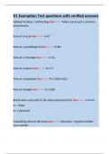
3.5 Prokaryotic Cells
(i) Know the ultrastructure of prokaryotic cells, including:
Structure Description Function
Cell wall Made of peptidoglycan (murein), not Provides strength and prevents lysis
cellulose
Capsule Outer polysaccharide layer (slimy) Protects from desiccation, immune system,
and antibiotics
Plasmid Small circular DNA molecule, separate from Carries genes for antibiotic resistance and
main DNA other traits
Flagellum Long protein tail, rotates Enables motility
Pili Short protein projections Used for attachment or conjugation (DNA
(fimbriae) transfer)
Ribosomes 70S type, smaller than eukaryotic 80S Site of protein synthesis
Circular DNA Single loop of DNA not bound in a nucleus Carries genetic information; floats free in
cytoplasm
Key Notes:
• No nucleus, no membrane-bound organelles.
• Some prokaryotes are capable of forming endospores under stress.
(ii) Understand the function of the structures listed above:
• Prokaryotic cells are adapted for survival in diverse environments.
• Capsule aids in pathogenicity (e.g. in Streptococcus pneumoniae).
• Plasmids allow for horizontal gene transfer and rapid evolution.
Topic 3: Cell Structure, Reproduction and Development
3.1 Know that all living organisms are made of cells, sharing some common features
Key Concept:
• The cell theory states that:
o All living organisms are composed of one or more cells.
o The cell is the basic structural and functional unit of life.
o All cells arise from pre-existing cells.
Shared Features of All Cells:
Feature Function
Plasma membrane Controls the movement of substances in and out of the cell
Cytoplasm Site of metabolic reactions
DNA Carries genetic information for controlling the cell
Ribosomes Site of protein synthesis
Enzymes Catalyse reactions essential to life
Energy release All cells must carry out respiration (aerobic or anaerobic) to release energy
Even though structure varies, these core features exist in both prokaryotic and eukaryotic cells.
3.2 Understand how the cells of multicellular organisms are organised into tissues, tissues into organs, and
organs into organ systems
Hierarchical Organisation in Multicellular Organisms:
1. Cells: The basic unit of life (e.g., epithelial cells, muscle cells).
2. Tissues: A group of similar cells working together to perform a specific function.
o Example: Muscle tissue contracts; epithelial tissue lines surfaces.
3. Organs: Made of different tissues working together to carry out a function.
o Example: The heart includes muscle tissue, nerve tissue, and connective tissue.
4. Organ Systems: Groups of organs that work together to carry out a broader function.
o Example: Circulatory system = heart + blood vessels; Digestive system = stomach, intestines,
liver, etc.
,Specialisation:
• Multicellular organisms rely on cell specialisation and cooperation for survival, as no single cell can
carry out all life functions.
3.3 Ultrastructure of Eukaryotic Cells
(i) Know the Ultrastructure:
Organelle Structure Function
Nucleus Surrounded by nuclear envelope with Stores genetic material; nucleolus
pores; contains DNA and nucleolus makes rRNA and ribosomes
Nucleolus Dense region inside nucleus Synthesises ribosomal RNA
Ribosomes Small, non-membranous; free or bound Site of protein synthesis
to rER
Rough Endoplasmic Membrane-bound sacs with ribosomes Synthesises and processes proteins for
Reticulum (rER) attached secretion or membrane use
Smooth ER Membrane-bound sacs without Lipid synthesis, detoxification
ribosomes
Mitochondria Double membrane, inner folds = cristae, Site of aerobic respiration (ATP
matrix inside production)
Golgi Apparatus Stacks of flattened sacs (cisternae), with Modifies, packages, and transports
vesicles proteins and lipids
, Lysosomes Vesicles containing hydrolytic enzymes Digests old organelles, pathogens, and
waste (intracellular digestion)
Centrioles Cylindrical structures made of Involved in spindle formation during
microtubules (in animal cells) cell division
Note: Only animal and plant cells are eukaryotic. Plant cells also include chloroplasts, a large permanent
vacuole, and a cellulose cell wall.
(ii) Understand Functions of Organelles:
• Protein synthesis and secretion involves coordination of nucleus → rER → Golgi → vesicle → plasma
membrane.
• Organelles provide compartmentalisation, increasing efficiency of metabolic processes.
• Mitochondria generate energy, while lysosomes manage cellular waste.
3.4 Role of rER and Golgi in Protein Transport and Enzyme Secretion
Protein Transport Pathway (with modifications and refinement):
1. Transcription in the nucleus – A gene coding for the protein is transcribed into mRNA.
2. mRNA exits the nucleus – The mRNA travels through nuclear pores into the cytoplasm.
3. Translation at ribosomes on the rough endoplasmic reticulum (rER) – Ribosomes on the rER synthesize
the polypeptide chain using the mRNA sequence.
4. Protein enters rER lumen – As the polypeptide is synthesized, it is threaded into the lumen of the rER.
5. Protein folding and modification in the rER:
o The polypeptide folds into its secondary and tertiary structures with the help of chaperone
proteins.
o Post-translational modifications may occur, such as the addition of carbohydrates to form
glycoproteins.
6. Packaging into transport vesicles – Properly folded and modified proteins are packaged into transport
vesicles that bud off from the rER.
7. Transport to the Golgi apparatus – Vesicles fuse with the cis face of the Golgi apparatus.
8. Further modification and sorting in the Golgi – The Golgi may carry out additional modifications (e.g.,
adding phosphate or sulfate groups) and sorts proteins based on their final destination.
9. Packaging into secretory vesicles – Modified proteins are packaged into secretory vesicles at the trans
face of the Golgi.
10. Exocytosis – Secretory vesicles move to the plasma membrane, fuse with it, and release the proteins
outside the cell by exocytosis.
, Formation of Extracellular Enzymes:
• Digestive enzymes (e.g., amylase, protease) are synthesised this way and secreted out of the cell to act
on substrates in the gut. (mentioned last page)
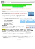
3.5 Prokaryotic Cells
(i) Know the ultrastructure of prokaryotic cells, including:
Structure Description Function
Cell wall Made of peptidoglycan (murein), not Provides strength and prevents lysis
cellulose
Capsule Outer polysaccharide layer (slimy) Protects from desiccation, immune system,
and antibiotics
Plasmid Small circular DNA molecule, separate from Carries genes for antibiotic resistance and
main DNA other traits
Flagellum Long protein tail, rotates Enables motility
Pili Short protein projections Used for attachment or conjugation (DNA
(fimbriae) transfer)
Ribosomes 70S type, smaller than eukaryotic 80S Site of protein synthesis
Circular DNA Single loop of DNA not bound in a nucleus Carries genetic information; floats free in
cytoplasm
Key Notes:
• No nucleus, no membrane-bound organelles.
• Some prokaryotes are capable of forming endospores under stress.
(ii) Understand the function of the structures listed above:
• Prokaryotic cells are adapted for survival in diverse environments.
• Capsule aids in pathogenicity (e.g. in Streptococcus pneumoniae).
• Plasmids allow for horizontal gene transfer and rapid evolution.