ASM 275: Lab 9 Questions And
Answers.
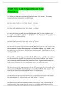
Q1. Click on the image area associated with the tooth crown. [10] - Answer The crown is
covered by the enamel toward the top of the image.
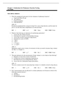
Q2. What types of teeth are these? [11] - Answer D. Molars
Q3. What tooth type is shown here? [12] - Answer A. Incisors
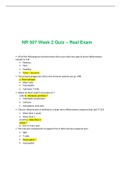
Q4. Select the area of the tooth associated with the crown. Note this tooth is broken in half
showing the enamel, dentin, and pulp chamber. [13] - Answer The crown is the top portion of
the tooth covered by white enamel.
Q5. What tooth type is shown here? [14] - Answer B. Canines
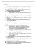
Q6. Select the area of this image associated with the LEFT canine, note this is the maxilla so the
sides are reversed. The right side of the image is the individual's left side. [15] - Answer from
the midline there are two incisors, then the canine, which is pointed. Please make sure you are
selecting the LEFT side.
Q7. Select the area of this image associated with the LEFT premolars, note this is the maxilla so
the sides are reversed. The right side of the image is the individual's left side. [15] - Answer
The premolars are oval in outline and have two primary cusps. They are located between the
single pointed canine and the multi-cusped molars toward the back of the mouth.
Q8. Select the area of this image associated with the LEFT molars, note this is the maxilla so the
sides are reversed. The right side of the image is the individual's left side. [15] - Answer The
molars square, multi-cusped and toward the back of the mouth (distal).
Q9. Which teeth are the first to begin forming (but just by a hair)? Look closely at the bottom
line associated with the 17th embryonic week. [18] - Answer A. Incisors
Q10. When the 2nd incisor has a NEARLY complete crown, the first molar is at what stage of
dental formation? Make sure to follow the correct line from left to right, beginning with the
Answers.
Q1. Click on the image area associated with the tooth crown. [10] - Answer The crown is
covered by the enamel toward the top of the image.
Q2. What types of teeth are these? [11] - Answer D. Molars
Q3. What tooth type is shown here? [12] - Answer A. Incisors
Q4. Select the area of the tooth associated with the crown. Note this tooth is broken in half
showing the enamel, dentin, and pulp chamber. [13] - Answer The crown is the top portion of
the tooth covered by white enamel.
Q5. What tooth type is shown here? [14] - Answer B. Canines
Q6. Select the area of this image associated with the LEFT canine, note this is the maxilla so the
sides are reversed. The right side of the image is the individual's left side. [15] - Answer from
the midline there are two incisors, then the canine, which is pointed. Please make sure you are
selecting the LEFT side.
Q7. Select the area of this image associated with the LEFT premolars, note this is the maxilla so
the sides are reversed. The right side of the image is the individual's left side. [15] - Answer
The premolars are oval in outline and have two primary cusps. They are located between the
single pointed canine and the multi-cusped molars toward the back of the mouth.
Q8. Select the area of this image associated with the LEFT molars, note this is the maxilla so the
sides are reversed. The right side of the image is the individual's left side. [15] - Answer The
molars square, multi-cusped and toward the back of the mouth (distal).
Q9. Which teeth are the first to begin forming (but just by a hair)? Look closely at the bottom
line associated with the 17th embryonic week. [18] - Answer A. Incisors
Q10. When the 2nd incisor has a NEARLY complete crown, the first molar is at what stage of
dental formation? Make sure to follow the correct line from left to right, beginning with the