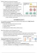
Autonomic reflexes are important for homeostasis.
- Sensory input information from somatosensory and
visceral receptors projects onto the homeostatic control
centers: hypothalamus, pons, and medulla.
- Hypothalamus also contains sensory neurons
(osmoreceptors and thermoreceptors)
- Alternatively, sensory information integrated within the
cerebral cortex and limbic system can produce
emotions that influence autonomic output.
- Motor output from the hypothalamus and brainstem
creates autonomic, endocrine, and behavioral responses.
- Some autonomic reflexes are capable of taking place without input from the brain – spinal reflexes.
- Urination, defecation, penile erection
AUTONOMIC NS (CHP 11)
The sympathetic and parasympathetic nervous system branches display all four of Walter Cannon’s
properties of homeostasis:
1. Preservation of the fitness of the internal environment.
2. Up-down regulation by tonic control.
3. Antagonistic control.
4. Chemical signals with different effects on different tissues.
Many internal organs are under (3) Antagonistic Control.
- One autonomic branch is excitatory the other is inhibitory.
- Effect often determined by neurotransmitter receptors on target tissue (e.g. adren-receptor on BVs).
- Exceptions to antagonistic control include sweat glands and smooth muscle in blood vessels.
Innervated only by the sympathetic branch.
Autonomic pathways have two efferent neurons in series. (1)
preganglionic neuron, (2) post ganglionic neuron.
- The (1) preganglionic neurons originate in the CNS and
project to an autonomic ganglion outside the CNS.
There, the preganglionic neuron projects onto the (2) postganglionic neuron.
- On average 1 preganglionic neuron diverges and projects onto 8-9 postganglionic neurons.
,
, Anatomical and Chemical Distinctions
The sympathetic and parasympathetic NS are anatomically distinct.
1. The pathway’s point of origin in the CNS:
a. Most sympathetic pathways originate in the thoracic and lumbar regions of the spinal cord..
b. Most parasympathetic pathways originate in the (1) brainstem or (2) sacral region of the SC.
i. (1) leaves the brain in cranial nerves, (2) leaves sacral region and controls pelvic organs
ii. Parasympathetic neurons project primarily to the head, neck, and internal organs
2. The location of the autonomic ganglia:
a. Sympathetic pathways: autonomic ganglia lie close to the SC, with short preganglionic neurons
and long postganglionic neurons.
b. Parasympathetic pathways: autonomic ganglia lie close to target organs, with long
preganglionic neurons and short postganglionic neurons.
- The parasympathetic system projects primarily (75%) via the vagus nerve (cranial nerve X)
The autonomic NS uses a number of chemical signals.
1. Both sympathetic and parasympathetic preganglionic neurons use acetylcholine (ACh) on nicotinic
cholinergic receptors (nAChR)
2. Most postganglionic sympathetic neurons secrete norepinephrine onto adrenergic receptors.
3. Most postganglionic parasympathetic neurons secrete acetylcholine on muscarinic cholinergic
receptors (mAChR)
Synapse between the postganglionic autonomic neuron and its target is called the neuroeffector junction.
- Autonomic neurons end with a postganglionic bulge or
varicosity, containing vesicles of NT.
- Underlying target tissue does not possess concentrated
regions of NT receptors.
- NT is released freely into the space, and
- Autonomic neurons simultaneously release a number of
other signalling molecules (e.g. histamine) which can inhibit
or stimulate the NT effect.
Neurotransmitter Release:
1. A depolarizing stimulus arrives at the varicosity
2. Voltage gated Ca2+ channels open
3. Ca2+ floods into the varicosity
4. Exocytosis of the vesicles is triggered, NT is released at the synapse.