HC 7 – general principles: diagnostic pathology
Kanker
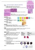
Invasie = cellen op plekken waar ze niet horen te zijn
Cytonucleaire atypie = grotere & lelijkere cellen
LVSI = invasie in lymfe- en bloedvaten = klomp cellen met ruimte eromheen
Organisatie vs geen organisatie
Paars = kanker
Roze = tumorcellen groeien sneller dan bloedvaten necrose
(tumorcellen die al dood zijn gegaan (geen kernen (paars))
Nomenclatuur
Mesenchymale tumoren Glad
Benigne: -oma Dwarsgestreept
Maligne: -sarcoma
Epitheliale tumoren
Benigne: -(cyst)adenoma / -papilloma
Maligne: -carcinoma
Melanocyten
Benigne: nervus
Maligne: melanoma
Diagnostiek
Diagnostiek = tumor of niet maligne of benigne
- Multidisciplinair team: verpleging, chirurgie, oncologen, radiologen & pathologen
Soms moeilijk kanker vaststellen: behandeling starten voor definitieve diagnose
Biopsie
Biopsie = beste manier om tumor te ontdekken (radiologie bv infectie) = fijne naald in tumor prikken
om kleine hoeveelheid materiaal te verkrijgen patholoog – ‘tissue is the issue’
- Makkelijk: borst & colon / grote tumor
- Moeilijk: pancreas & long / kleine tumor
Benigne vs maligne
Maligne = invasie + cytonucleaire atypie + metastase (LVSI)
- Invasie = geen herkenning van anatomische grenzen
- Metastase = naar bloed & lymfevaten
Pre-maligne = (nog) geen invasie + cytonucleaire atypie
Benigne = geen invasie + geen cytonucleaire atypie
Normaal
Georganiseerd
Duidelijke begrenzing van cellen en weefsels
Regelmatige cellen (max 1,5 x erythrocyt)
Dezelfde grootte en vorm
Maligniteit (cytonucleaire atypia)
Groot & variabel gevormd nuclei met hyperchromasie & polychromasie (donkere kern)
Veel delende cellen
Ongeorganiseerd
Polymorfisme/pleomorfisme = variatie in grootte & vorm
Verlies normale celkenmerken
Borsten
,Normale anatomie: lumen, luminale epitheelcellen (productie melk) & myoepitheelcellen
(samentrekken om melk te transporteren)
- DCIS = ductus carcinoom in situ = hyperplasie epitheliale cellen maar in ductus
- Adenocarcinoom = epitheliale cellen invasie myoepitheel
staining: myoepitheel (grens) intact of niet (bruin)
Huid
Gekeratiniseerd plaveiselepitheel: onder stamceldeling & boven afgeplat epitheel
Squamous cell carcinoom = grote ronde cellen aan de bovenkant van het epitheel
Waarom doen we een biopsie?
Nadelen: invasief, pijnlijk, risico’s (bloeden/pneumothorax(long)/ent-metastase (sarcoma)/infectie)
Voordelen:
Diagnose: wat voor tumor?
Prognose
Biomarker analyse
o Prognotisch: hoe groot is de kans op agressief gedrag (grading)
o Therapeutisch: hoe groot is de kans op reactie op behandelingen
Grading
Grading = abnormaliteit cellen/aggressiviteit tumor bepalen – waarschijnlijkheid verspreiding?
Grade I: differentiatie Grade 4: anaplasie
Staging = T (grootte & uitbreiding), N (lymfeknopen) & M (metastasen) – verspreiding & grootte?
Stage 0: in situ Stage IV: metastase
Prognose & behandeling (kankerbehandeling pas al tumor ook schade levert)
We zijn hier niet goed in
Immuunhistochemie
Immuunhistochemie = weefsel met antigenen monoklonale primaire antlichamen secundaire
antlichamen (met tag) fluorescentie
Borstkanker:
Oestrogeenreceptoren bruin
HER2-receptoren: tumor amplificeren groeipotentie
Chemotherapie: anti-HER2 antistoffen blokkeren HER2-receptoren 80% elimineren
Vragen
, B Geen 100% Yes
B C A, C & D
A A (cyst = bol met vloeistof)
A (adeno = klieren (mucus) & squamous = huid/oesophagus: keratine)
C (veel mitose)
B?
HC 8 – general principles: molecular diagnostics
Kanker
Invasie = cellen op plekken waar ze niet horen te zijn
Cytonucleaire atypie = grotere & lelijkere cellen
LVSI = invasie in lymfe- en bloedvaten = klomp cellen met ruimte eromheen
Organisatie vs geen organisatie
Paars = kanker
Roze = tumorcellen groeien sneller dan bloedvaten necrose
(tumorcellen die al dood zijn gegaan (geen kernen (paars))
Nomenclatuur
Mesenchymale tumoren Glad
Benigne: -oma Dwarsgestreept
Maligne: -sarcoma
Epitheliale tumoren
Benigne: -(cyst)adenoma / -papilloma
Maligne: -carcinoma
Melanocyten
Benigne: nervus
Maligne: melanoma
Diagnostiek
Diagnostiek = tumor of niet maligne of benigne
- Multidisciplinair team: verpleging, chirurgie, oncologen, radiologen & pathologen
Soms moeilijk kanker vaststellen: behandeling starten voor definitieve diagnose
Biopsie
Biopsie = beste manier om tumor te ontdekken (radiologie bv infectie) = fijne naald in tumor prikken
om kleine hoeveelheid materiaal te verkrijgen patholoog – ‘tissue is the issue’
- Makkelijk: borst & colon / grote tumor
- Moeilijk: pancreas & long / kleine tumor
Benigne vs maligne
Maligne = invasie + cytonucleaire atypie + metastase (LVSI)
- Invasie = geen herkenning van anatomische grenzen
- Metastase = naar bloed & lymfevaten
Pre-maligne = (nog) geen invasie + cytonucleaire atypie
Benigne = geen invasie + geen cytonucleaire atypie
Normaal
Georganiseerd
Duidelijke begrenzing van cellen en weefsels
Regelmatige cellen (max 1,5 x erythrocyt)
Dezelfde grootte en vorm
Maligniteit (cytonucleaire atypia)
Groot & variabel gevormd nuclei met hyperchromasie & polychromasie (donkere kern)
Veel delende cellen
Ongeorganiseerd
Polymorfisme/pleomorfisme = variatie in grootte & vorm
Verlies normale celkenmerken
Borsten
,Normale anatomie: lumen, luminale epitheelcellen (productie melk) & myoepitheelcellen
(samentrekken om melk te transporteren)
- DCIS = ductus carcinoom in situ = hyperplasie epitheliale cellen maar in ductus
- Adenocarcinoom = epitheliale cellen invasie myoepitheel
staining: myoepitheel (grens) intact of niet (bruin)
Huid
Gekeratiniseerd plaveiselepitheel: onder stamceldeling & boven afgeplat epitheel
Squamous cell carcinoom = grote ronde cellen aan de bovenkant van het epitheel
Waarom doen we een biopsie?
Nadelen: invasief, pijnlijk, risico’s (bloeden/pneumothorax(long)/ent-metastase (sarcoma)/infectie)
Voordelen:
Diagnose: wat voor tumor?
Prognose
Biomarker analyse
o Prognotisch: hoe groot is de kans op agressief gedrag (grading)
o Therapeutisch: hoe groot is de kans op reactie op behandelingen
Grading
Grading = abnormaliteit cellen/aggressiviteit tumor bepalen – waarschijnlijkheid verspreiding?
Grade I: differentiatie Grade 4: anaplasie
Staging = T (grootte & uitbreiding), N (lymfeknopen) & M (metastasen) – verspreiding & grootte?
Stage 0: in situ Stage IV: metastase
Prognose & behandeling (kankerbehandeling pas al tumor ook schade levert)
We zijn hier niet goed in
Immuunhistochemie
Immuunhistochemie = weefsel met antigenen monoklonale primaire antlichamen secundaire
antlichamen (met tag) fluorescentie
Borstkanker:
Oestrogeenreceptoren bruin
HER2-receptoren: tumor amplificeren groeipotentie
Chemotherapie: anti-HER2 antistoffen blokkeren HER2-receptoren 80% elimineren
Vragen
, B Geen 100% Yes
B C A, C & D
A A (cyst = bol met vloeistof)
A (adeno = klieren (mucus) & squamous = huid/oesophagus: keratine)
C (veel mitose)
B?
HC 8 – general principles: molecular diagnostics