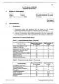
Chapter 17
Cardiovascular
Normal Heart Anatomy & Function (pg 553-555)
Normal Blood Flow:
1. Deoxygenated blood returns from the body to the right atrium via the superior and inferior vena
cava.
2. Blood flows through the tricuspid valve into the right ventricle.
3. Upon contraction, the right ventricle pumps blood through the pulmonary valve into the
pulmonary arteries, leading to the lungs for oxygenation.
4. Oxygenated blood returns to the left atrium via the pulmonary veins.
5. Blood passes through the mitral (bicuspid) valve into the left ventricle.
6. The left ventricle contracts, sending blood through the aortic valve into the aorta, distributing
oxygen-rich blood to the body.
Define Cardiac Output: Cardiac Output (CO) is the volume of blood the heart pumps per minute. It is
calculated by multiplying the heart rate (HR) by the stroke volume (SV): CO = HR × SV. This
measurement indicates the efficiency of the heart's pumping ability and overall circulatory health.
, Congestive Heart Failure (CHF) (pgs 556-557, 560)
Brief Pathophysiology: CHF occurs when the heart cannot pump blood effectively to meet the body's
needs, leading to inadequate oxygen and nutrient delivery to tissues. This inefficiency causes blood to
back up into the lungs and other parts of the body, resulting in congestion and fluid accumulation.
Signs & Symptoms/Assessment Findings:
o Infants: Poor feeding, tachypnea, diaphoresis during feeding, poor weight gain.
o Children: Exercise intolerance, fatigue, weight gain due to fluid retention, dyspnea, edema,
hepatomegaly.
Diagnostic Tests:
o Chest X-ray: May reveal cardiomegaly and pulmonary congestion.
o Echocardiogram: Assesses heart structure and function, identifying underlying causes.
o Electrocardiogram (ECG): Detects arrhythmias and ventricular hypertrophy.
o Laboratory Tests: Brain Natriuretic Peptide (BNP) levels can indicate heart failure severity.
Treatment:
o Nursing Care:
Monitor vital signs, fluid balance, and weight.
Administer prescribed medications.
Provide oxygen therapy as needed.
Educate caregivers on symptom management and medication adherence.
o Medical Care:
Diuretics to reduce fluid overload.
Inotropes to improve cardiac contractility.
Afterload-reducing agents (e.g., ACE inhibitors) to decrease cardiac workload.
Education:
o Teach families about medication administration and potential side effects.
o Emphasize the importance of regular follow-up appointments.
o Provide guidance on recognizing signs of worsening heart failure, such as increased shortness of
breath or edema.
Prevention/Screening of Congenital Heart Disease (CHD) (pg 555-556)
Prevention:
o Encourage maternal health optimization before and during pregnancy, including managing
chronic conditions and avoiding teratogenic substances.