Microbiology Midterm Study Guide
Microbiology Midterm Study Guide
questions And Answers
Covers Chapters 1-13 /Week 1 (Chapters 1-3)
v v v v v v
1. Pasteur - Final disproof spontaneous generation
v v v v v
2. Hook- Saw first microbes v v v
3. Lister -Aseptic techniques during surgery
v v v v
4. Semmelweis - Dr. had to wash hands in maternity ward v v v v v v v v v
5. Schultze & Schwann- Chemical treatment of air stops ability to produce life
v v v v v v v v v v v
6. Koch - Developed postulates for disease microbe connection
v v v v v v v
7. Redi - Maggot-meat experiment
v v v
8. Leeuwenhoek - Made first microscope v v v v
Fundamental of cells: v v
Unicellular (Bacteria, Archaea, Protozoa, some fungi) and multicellular organisms (animals and plants)
v v v v v v v v v v v
▪ All cells (prokaryotes + eukaryotes) have in common:
v v v v v v v
Cell membrane v
DNAthat holds genetic information v v v v
Ribosomes for protein synthesis v v v v
Cytoplasm v
Eukaryotes are more complex with DNA enclosed in nucleus and membrane enclosed organelles
v v v v v v v v v v v v
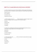
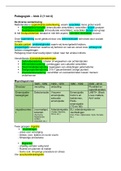
Bacterial shapes:
v
Cocci Rods. Vibrio Spirillum
1
, Microbiology Midterm Study Guide
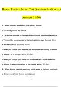
Spirochete Branching Filaments
v
2
, Microbiology Midterm Study Guide
The 5 Is:
v v
Inoculation - Purposely moving something from 1 place to another. starts with specimen collection; lesion,
v v v v v v v v v v v v v v
draw blood, bird droppings, etc.
v v v v v
- introducing a tiny sample into a medium to provide an env't where they multiply.
v v v v v v v v v v v v v
Incubation - To hear bacteria to make it grow (usually body temp). maintaining something at the most
v v v v v v v v v v v v v v v v
favorable temperature for its development. 20 deg C & 40 deg. C02 may be required.
v v v v v v v v v v v v v v v
- promotes multiplication of microbes over period of hours.
v v v v v v v
- produces a culture v v
Isolation - Separate from each other
v v v v v
separation of a strain from a natural, mixed population of living microbes, spreading bacteria apart as far as
v v v v v v v v v v v v v v v v v
possible.
v
- isolated microbes takes the form of separate colonies on solid media or turbidity (free floating cells) on
v v v v v v v v v v v v v v v v
broth.
v
Inspection - Doing tests on the bug v v v v v v
appearance, cells, colony (red? shape, gram stain, sugar, etc.).
v v v v v v v v
Identification - determine type of microbe v v v v v
- specialized tests; biochemical test to determine metabolic activities specific to microbes
v v v v v v v v v v
- immunologic tests, genetic analysis. v v v
Microscopy Basics/Types of microscopes: v v v
1. Bright field microscope - Most widely used microscope v v v v v v v
image is darker than illuminated field
v v v v v v
made by putting light through specimen v v v v v
2. Dark field microscope- similar to bright field
v v v v v v
image is lighter than illuminated field
v v v v v v
bright field microscope is changed to dark field microscope by adding a condenser to the light
v v v v v v v v v v v v v v v
3. Phase contrast microscope- used with live specimen v v v v v v
produces image with specimen against gray background
v v v v v v v
can see internal cells
v v v v
4. Differential interreference microscope- produces colorful 3D image 2 v v v v v v v
prisms which add contrasting colors to image
v v v v v v v
3
, Microbiology Midterm Study Guide
5. Fluorescence microscope – ultraviolet light for colored image against black field v v v v v v v v v v
makes an image
v v v
6. Confocal microscope- visualizes fluorescent molecules in a single plane of focus by excluding
v v v v v v v v v v v v
out of focus light
v v v v
7. Transmission electron microscope- uses electron beams to form image v v v v v v v v
magnify images up to 100,000x
v v v v v
works by transmitting electrons through specimen
v v v v v
8. Scanning electron microscope – detailed 3D images of specimen v v v v v v v v
magnify up to 650,000x
v v v v
works by sending electrons to the specimen and detecting deflected electrons is characterized as a
v v v v v v v v v v v v v v
microscope that has lower magnifying power but can provide 3-dimensional viewing of objects.
v v v v v v v v v v v v v
captures the image of the object in black and white after being stained with gold and palladium.
v v v v v v v v v v v v v v v v v
9. Atomic force microscope- A microscope that uses a beam deflection system with a laser and
v v v v v v v v v v v v v v
photodetector to measure the beam position as a cantilever with a tip scans across the surface of a
v v v v v v v v v v v v v v v v v v
material. The force between the tip and sample is calculated by measuring the deflection of the
v v v v v v v v v v v v v v v v
lever and knowing the stiffness of the cantilever.
v v v v v v v v
10. Scanning tunneling microscope- uses a computer to make highly magnified image of a
v v v v v v v v v v v v
specimenv
Advantage: can view other objects as small as an individual atom, can view living organisms
v v v v v v v v v v v v v v
Disadvantage: expensive
v v
Light microscopes Vs Electron microscopes:
v v v v v
1. light microscope uses light to illuminate specimens and glass lenses to magnify images.
v v v v v v v v v v v v
2. electron microscope uses a beam of electrons to illuminate specimens and magnetic lenses to
v v v v v v v v v v v v v
magnify images. The resolution (the level of image detailing) is the main difference between these
v v v v v v v v v v v v v v v
two microscopes
v v
Resolution:
• A scanning transmission electron microscope has achieved better than 50 pm resolution in
v v v v v v v v v v v v
annular dark-field imaging mode and magnifications of up to about 10,000,000×
v v v v v v v v v v v
• light microscopes are limited by diffraction to about 200 nm resolution and useful magnifications
v v v v v v v v v v v v v
below 2000×.
v v
Magnification:
• Magnification is the ability to make small objects seem larger, such as making v v v v v v v v v v v v
a microscopic organism visible. Resolution is the ability to distinguish two objects from each other.
v v v v v v v v v v v v v v
Light microscopy has limits to both its resolution and its magnification.
v v v v v v v v v v v
Macromolecules key to microbiology: v v v
Macromolecules: Large organic molecules in cells. Includes nucleic acids, proteins, polysaccharides and
v v v v v v v v v v v
lipids
v
polysaccharide - is one type of macromolecule made up of monosaccharides like glucose and fructose
v v v v v v v v v v v v v v
4
Microbiology Midterm Study Guide
questions And Answers
Covers Chapters 1-13 /Week 1 (Chapters 1-3)
v v v v v v
1. Pasteur - Final disproof spontaneous generation
v v v v v
2. Hook- Saw first microbes v v v
3. Lister -Aseptic techniques during surgery
v v v v
4. Semmelweis - Dr. had to wash hands in maternity ward v v v v v v v v v
5. Schultze & Schwann- Chemical treatment of air stops ability to produce life
v v v v v v v v v v v
6. Koch - Developed postulates for disease microbe connection
v v v v v v v
7. Redi - Maggot-meat experiment
v v v
8. Leeuwenhoek - Made first microscope v v v v
Fundamental of cells: v v
Unicellular (Bacteria, Archaea, Protozoa, some fungi) and multicellular organisms (animals and plants)
v v v v v v v v v v v
▪ All cells (prokaryotes + eukaryotes) have in common:
v v v v v v v
Cell membrane v
DNAthat holds genetic information v v v v
Ribosomes for protein synthesis v v v v
Cytoplasm v
Eukaryotes are more complex with DNA enclosed in nucleus and membrane enclosed organelles
v v v v v v v v v v v v
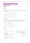
Bacterial shapes:
v
Cocci Rods. Vibrio Spirillum
1
, Microbiology Midterm Study Guide
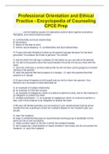
Spirochete Branching Filaments
v
2
, Microbiology Midterm Study Guide
The 5 Is:
v v
Inoculation - Purposely moving something from 1 place to another. starts with specimen collection; lesion,
v v v v v v v v v v v v v v
draw blood, bird droppings, etc.
v v v v v
- introducing a tiny sample into a medium to provide an env't where they multiply.
v v v v v v v v v v v v v
Incubation - To hear bacteria to make it grow (usually body temp). maintaining something at the most
v v v v v v v v v v v v v v v v
favorable temperature for its development. 20 deg C & 40 deg. C02 may be required.
v v v v v v v v v v v v v v v
- promotes multiplication of microbes over period of hours.
v v v v v v v
- produces a culture v v
Isolation - Separate from each other
v v v v v
separation of a strain from a natural, mixed population of living microbes, spreading bacteria apart as far as
v v v v v v v v v v v v v v v v v
possible.
v
- isolated microbes takes the form of separate colonies on solid media or turbidity (free floating cells) on
v v v v v v v v v v v v v v v v
broth.
v
Inspection - Doing tests on the bug v v v v v v
appearance, cells, colony (red? shape, gram stain, sugar, etc.).
v v v v v v v v
Identification - determine type of microbe v v v v v
- specialized tests; biochemical test to determine metabolic activities specific to microbes
v v v v v v v v v v
- immunologic tests, genetic analysis. v v v
Microscopy Basics/Types of microscopes: v v v
1. Bright field microscope - Most widely used microscope v v v v v v v
image is darker than illuminated field
v v v v v v
made by putting light through specimen v v v v v
2. Dark field microscope- similar to bright field
v v v v v v
image is lighter than illuminated field
v v v v v v
bright field microscope is changed to dark field microscope by adding a condenser to the light
v v v v v v v v v v v v v v v
3. Phase contrast microscope- used with live specimen v v v v v v
produces image with specimen against gray background
v v v v v v v
can see internal cells
v v v v
4. Differential interreference microscope- produces colorful 3D image 2 v v v v v v v
prisms which add contrasting colors to image
v v v v v v v
3
, Microbiology Midterm Study Guide
5. Fluorescence microscope – ultraviolet light for colored image against black field v v v v v v v v v v
makes an image
v v v
6. Confocal microscope- visualizes fluorescent molecules in a single plane of focus by excluding
v v v v v v v v v v v v
out of focus light
v v v v
7. Transmission electron microscope- uses electron beams to form image v v v v v v v v
magnify images up to 100,000x
v v v v v
works by transmitting electrons through specimen
v v v v v
8. Scanning electron microscope – detailed 3D images of specimen v v v v v v v v
magnify up to 650,000x
v v v v
works by sending electrons to the specimen and detecting deflected electrons is characterized as a
v v v v v v v v v v v v v v
microscope that has lower magnifying power but can provide 3-dimensional viewing of objects.
v v v v v v v v v v v v v
captures the image of the object in black and white after being stained with gold and palladium.
v v v v v v v v v v v v v v v v v
9. Atomic force microscope- A microscope that uses a beam deflection system with a laser and
v v v v v v v v v v v v v v
photodetector to measure the beam position as a cantilever with a tip scans across the surface of a
v v v v v v v v v v v v v v v v v v
material. The force between the tip and sample is calculated by measuring the deflection of the
v v v v v v v v v v v v v v v v
lever and knowing the stiffness of the cantilever.
v v v v v v v v
10. Scanning tunneling microscope- uses a computer to make highly magnified image of a
v v v v v v v v v v v v
specimenv
Advantage: can view other objects as small as an individual atom, can view living organisms
v v v v v v v v v v v v v v
Disadvantage: expensive
v v
Light microscopes Vs Electron microscopes:
v v v v v
1. light microscope uses light to illuminate specimens and glass lenses to magnify images.
v v v v v v v v v v v v
2. electron microscope uses a beam of electrons to illuminate specimens and magnetic lenses to
v v v v v v v v v v v v v
magnify images. The resolution (the level of image detailing) is the main difference between these
v v v v v v v v v v v v v v v
two microscopes
v v
Resolution:
• A scanning transmission electron microscope has achieved better than 50 pm resolution in
v v v v v v v v v v v v
annular dark-field imaging mode and magnifications of up to about 10,000,000×
v v v v v v v v v v v
• light microscopes are limited by diffraction to about 200 nm resolution and useful magnifications
v v v v v v v v v v v v v
below 2000×.
v v
Magnification:
• Magnification is the ability to make small objects seem larger, such as making v v v v v v v v v v v v
a microscopic organism visible. Resolution is the ability to distinguish two objects from each other.
v v v v v v v v v v v v v v
Light microscopy has limits to both its resolution and its magnification.
v v v v v v v v v v v
Macromolecules key to microbiology: v v v
Macromolecules: Large organic molecules in cells. Includes nucleic acids, proteins, polysaccharides and
v v v v v v v v v v v
lipids
v
polysaccharide - is one type of macromolecule made up of monosaccharides like glucose and fructose
v v v v v v v v v v v v v v
4