Would advise to
on full screen to
make ECGs look
☺
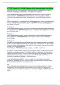
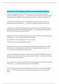
ECG
, The ECG wave
oP Wave = atrial depol (0.08-0.10 seconds
big box
oPR Interval = conduction through AV nod
(0.12-0.2 seconds) ≤ 1 big box
oQRS complex = ventricular depol (0.06-0
seconds) ~ ≤ 0.5 big box
oST segment = time between ventricular d
and repol
oT Wave = ventricular repol
oQT Interval = time taken for ventricular d
This Photo by Unknown Author is licensed under CC BY-SA
repol (0.2-0.4seconds) ~1-2 big boxes
,
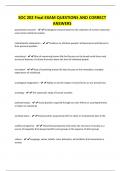
, What is Axis?
• Axis is the direction of electrical conduction
the heart.
• Normal conduction
• SA Node → AV node→ His Purkinje System
3. Axis ventricular contraction
• Normal cardiac axis lies between -30- +90
degrees
• Most positive deflections are in leads I, II an
as electrical activity flows towards these lea
• Most negative deflection in lead aVR as this
lead looks at heart in opposite direction
on full screen to
make ECGs look
☺
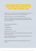
ECG
, The ECG wave
oP Wave = atrial depol (0.08-0.10 seconds
big box
oPR Interval = conduction through AV nod
(0.12-0.2 seconds) ≤ 1 big box
oQRS complex = ventricular depol (0.06-0
seconds) ~ ≤ 0.5 big box
oST segment = time between ventricular d
and repol
oT Wave = ventricular repol
oQT Interval = time taken for ventricular d
This Photo by Unknown Author is licensed under CC BY-SA
repol (0.2-0.4seconds) ~1-2 big boxes
,
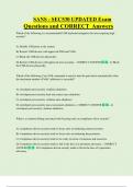
, What is Axis?
• Axis is the direction of electrical conduction
the heart.
• Normal conduction
• SA Node → AV node→ His Purkinje System
3. Axis ventricular contraction
• Normal cardiac axis lies between -30- +90
degrees
• Most positive deflections are in leads I, II an
as electrical activity flows towards these lea
• Most negative deflection in lead aVR as this
lead looks at heart in opposite direction