Muscular System
Overview of the Muscular System
• Developmental Origin:
o The muscular system develops from the mesodermal germ layer and comprises
three types of muscle:
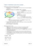
1. Skeletal Muscle: Derived primarily from paraxial mesoderm, which forms
somites (occipital to sacral regions) and somitomeres in the head. This
includes limb and body wall muscles.
2. Smooth Muscle: Mainly arises from visceral splanchnic mesoderm
surrounding the gut and its derivatives. Smooth muscle of pupillary,
mammary gland, and sweat gland muscles comes from ectoderm.
3. Cardiac Muscle: Originates from visceral splanchnic mesoderm
surrounding the heart tube, giving rise to the myocardium and contributing
to the heart's contractile ability.
Development of Skeletal Muscle
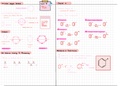
Formation from Somites
• Somites and Somitomeres:
o Head Musculature: Develops from seven somitomeres (derived from paraxial
mesoderm). These partially segmented mesenchymal whorls give rise to the muscles
of the head (except muscles of the iris).
o Musculature of the Axial Skeleton, Body Wall, and Limbs:
§ Derived from somites, which initially form as somitomeres and extend from
the occipital region to the tail bud.
§ These somites give rise to skeletal muscles of the back, body wall, and
limbs.
• Somite Differentiation:
o Epithelization:
§ Immediately after segmentation, somitomeres undergo epithelization, forming
a ball of epithelial cells around a central cavity.
o Sclerotome and Dermomyotome Formation:
§ The ventral region of each somite becomes mesenchymal, forming the
sclerotome. The sclerotome differentiates into the bone-forming cells for
the vertebrae and ribs.
§ The upper region of the somite forms the dermatome and two muscle-
forming areas: the ventrolateral (VLL) and dorsomedial (DML) lips. These
, muscle precursors form progenitor muscle cells that aggregate to form the
dermomyotome.
• Muscle Progenitors and Migration:
o Cells from the VLL:
§ Some cells migrate into the parietal layer of the lateral plate mesoderm,
forming muscles such as the infrahyoid muscles, abdominal wall muscles
(including rectus abdominis, internal/external oblique, and transversus
abdominis), and limb muscles.
o Cells from the DML and Remaining VLL:
§ Contribute to muscles of the back, shoulder girdle, and intercostal
muscles.
Lateral Somitic Frontier
• Definition:
o The lateral somitic frontier is the boundary between the somite-derived mesoderm
and the parietal layer of lateral plate mesoderm.
o It separates two key mesodermal domains:
1. Primaxial Domain: Contains only somite-derived cells from paraxial
mesoderm and is found around the neural tube.
2. Abaxial Domain: Contains cells from both the parietal layer of lateral plate
mesoderm and migratory somite cells that cross the lateral somitic frontier.
• Muscle Differentiation Based on Domains:
o Primaxial Muscle Cells:
§ Include back muscles, intercostals, and deep muscles of the neck.
§ Receive signals from the neural tube and notochord for their differentiation.
o Abaxial Muscle Cells:
§ Include abdominal muscles, limb muscles, and infrahyoid muscles.
§ Differentiate under signals primarily from the lateral plate mesoderm.
Innervation of Skeletal Muscles
• Nervous System Contributions:
o The new classification of muscle development relies on whether muscle precursors
belong to the primaxial or abaxial domains, rather than the older concepts of
epimeres (back muscles) and hypomeres (limb and body wall muscles).
o Primaxial Muscles are generally innervated by dorsal primary rami.
o Abaxial Muscles are innervated by ventral primary rami.
Overview of the Muscular System
• Developmental Origin:
o The muscular system develops from the mesodermal germ layer and comprises
three types of muscle:
1. Skeletal Muscle: Derived primarily from paraxial mesoderm, which forms
somites (occipital to sacral regions) and somitomeres in the head. This
includes limb and body wall muscles.
2. Smooth Muscle: Mainly arises from visceral splanchnic mesoderm
surrounding the gut and its derivatives. Smooth muscle of pupillary,
mammary gland, and sweat gland muscles comes from ectoderm.
3. Cardiac Muscle: Originates from visceral splanchnic mesoderm
surrounding the heart tube, giving rise to the myocardium and contributing
to the heart's contractile ability.
Development of Skeletal Muscle
Formation from Somites
• Somites and Somitomeres:
o Head Musculature: Develops from seven somitomeres (derived from paraxial
mesoderm). These partially segmented mesenchymal whorls give rise to the muscles
of the head (except muscles of the iris).
o Musculature of the Axial Skeleton, Body Wall, and Limbs:
§ Derived from somites, which initially form as somitomeres and extend from
the occipital region to the tail bud.
§ These somites give rise to skeletal muscles of the back, body wall, and
limbs.
• Somite Differentiation:
o Epithelization:
§ Immediately after segmentation, somitomeres undergo epithelization, forming
a ball of epithelial cells around a central cavity.
o Sclerotome and Dermomyotome Formation:
§ The ventral region of each somite becomes mesenchymal, forming the
sclerotome. The sclerotome differentiates into the bone-forming cells for
the vertebrae and ribs.
§ The upper region of the somite forms the dermatome and two muscle-
forming areas: the ventrolateral (VLL) and dorsomedial (DML) lips. These
, muscle precursors form progenitor muscle cells that aggregate to form the
dermomyotome.
• Muscle Progenitors and Migration:
o Cells from the VLL:
§ Some cells migrate into the parietal layer of the lateral plate mesoderm,
forming muscles such as the infrahyoid muscles, abdominal wall muscles
(including rectus abdominis, internal/external oblique, and transversus
abdominis), and limb muscles.
o Cells from the DML and Remaining VLL:
§ Contribute to muscles of the back, shoulder girdle, and intercostal
muscles.
Lateral Somitic Frontier
• Definition:
o The lateral somitic frontier is the boundary between the somite-derived mesoderm
and the parietal layer of lateral plate mesoderm.
o It separates two key mesodermal domains:
1. Primaxial Domain: Contains only somite-derived cells from paraxial
mesoderm and is found around the neural tube.
2. Abaxial Domain: Contains cells from both the parietal layer of lateral plate
mesoderm and migratory somite cells that cross the lateral somitic frontier.
• Muscle Differentiation Based on Domains:
o Primaxial Muscle Cells:
§ Include back muscles, intercostals, and deep muscles of the neck.
§ Receive signals from the neural tube and notochord for their differentiation.
o Abaxial Muscle Cells:
§ Include abdominal muscles, limb muscles, and infrahyoid muscles.
§ Differentiate under signals primarily from the lateral plate mesoderm.
Innervation of Skeletal Muscles
• Nervous System Contributions:
o The new classification of muscle development relies on whether muscle precursors
belong to the primaxial or abaxial domains, rather than the older concepts of
epimeres (back muscles) and hypomeres (limb and body wall muscles).
o Primaxial Muscles are generally innervated by dorsal primary rami.
o Abaxial Muscles are innervated by ventral primary rami.