3.6 Neuropsychology
Lecture 1 - Introduction
Learning goals
● To learn the organisation of the brain and the brain anatomy of the parietal and occipital lobes as well
as the basal ganglia
● To understand how motion and perception are processed in the brain
● To link lesions of the parietal and occipital lobes to motion and perception disorders
Neuropsychology
● A branch of psychology
● The study of the brain mechanisms in relation to cognitive and behavioral processes
● Modern imaging techniques allow a huge progress in the knowledge of our brain
Neurological damage
Brain damage has long provided insights to the relation between brain and behavior
1. Vascular disorders (stroke): sudden onset cerebrovascular events
● Cerebral hemorrhage: bleeding in the brain
● Cerebral ischemia: disruption of blood supply (thrombosis, embolism, arteriosclerosis)
2. Tumors: a mass of cells, which grows independently of the rest of the body
3. Trauma: injuries of the brain (close- or open-head)
4. Epilepsy: excessive and abnormal pattern of activity in the brain, it induces a transient loss of
consciousness
5. Degenerative disorders: entails the death of neurons, motor or cognitive difficulties often noticed by
family members
● Parkinson disease: degeneration of the basal ganglia, resulting in reduction of the dopamine
and difficulties in initiating a behavior
● Alzheimer disease: degeneration of the neurons in the cortex, characterized by attentional
deficits, forgiveness, changes in personality
● Huntington disease: genetic disease, which involves atrophy of the striatum. It first impairs
motoric abilities and afterwards cognitive processes
● Korsakoff disease: degeneration of the diencephalon, mainly due to chronic alcoholism and
malnutrition, it produces amnesia
● Multiple sclerosis: degeneration of the myelin surrounding the axons, which can lead to
degeneration of the neuron itself, it is an autoimmune disorder
Research methods
1. Psychophysiological indices: measure the peripheral responses
● Electrodermal activity (EDA) → skin conductance measure, “sweat response”
○ Measure the activation of the sympathetic system meaning the
physiological arousal (activation) necessary to initiate a behavioral
response
○ Is larger to both positive (e.g., cute picture bear) and negative (e.g.,
snake) pictures as compared to neutral ones
● ELectromyogram (EMG)
● Electrocardiogram (ECG)
1
, 2. Imaging techniques: measure the brain activation
● Electro-encephalogram (EEG)
○ Measures the electrical signal originating from the pyramidal cells, which
has a negative charge
○ When numerous pyramidal cells are activated synchronically, we can
detect relatively large waves on the scalp, which are called event-related
potentials (ERPs)
● (Functional) magnet resonance imaging (fMRI and MRI)
○ fMRI measures the metabolic signal originating from the blood oxygen
level
○ Blood-oxygen level-dependent (BOLD) signal is an indirect index of neural
activation
● Positron emission tomography (PET)
Anatomy of cognition
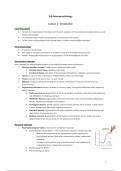
Anatomical organization
● Rostral/Anterior: parts of the body toward the nose
● Caudal/Posterior: parts of the body toward the tail
● Dorsal: parts of the body pointing up from the back
● Ventral: parts of the body pointing down from the belly
● Medial: structures close to the midline
● Lateral: structures far from the midline
○ Ipsilateral: structures on the same side of the body
○ Contralateral: structures on the opposite side of the body
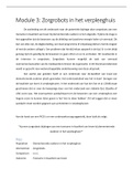
Anatomical Nervous System Divisions
2
, Nervous System
1. Central nervous system (CNS): processes and integrates information, coordinating bodily functions
and responses
● Brain
● Spinal cord
2. Peripheral nervous system (PNS): connects the central nervous system to the rest of the body,
enabling communication between the brain, spinal cord, and limbs/organs
● Somatic nervous system: controls voluntary movements and transmits sensory information
(e.g., vision, hearing, touch) to the central nervous system
● Autonomic nervous system: regulates involuntary physiological processes (e.g., heart rate,
digestion, and responses to stimuli like stress or relaxation)
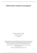
Anatomical organization
1. Myelencephalon (or medulla): connects
the brain with the spinal cord
2. Metencephalon (or hindbrain): consists
of pons and cerebellum, somatosensory
structure
3. Mesencephalon (or midbrain): consists
of tegmentum, periaqueductal grey
(PAG), visuomotor structure
4. Diencephalon: consists of
hypothalamus and thalamus, referred as
“gateway to the cortex”
5. Telencephalon: consists of the cerebral
cortex (i.e., neocortex) and sub-cortical
structures (i.e., basal ganglia, limbic
system)
3
Lecture 1 - Introduction
Learning goals
● To learn the organisation of the brain and the brain anatomy of the parietal and occipital lobes as well
as the basal ganglia
● To understand how motion and perception are processed in the brain
● To link lesions of the parietal and occipital lobes to motion and perception disorders
Neuropsychology
● A branch of psychology
● The study of the brain mechanisms in relation to cognitive and behavioral processes
● Modern imaging techniques allow a huge progress in the knowledge of our brain
Neurological damage
Brain damage has long provided insights to the relation between brain and behavior
1. Vascular disorders (stroke): sudden onset cerebrovascular events
● Cerebral hemorrhage: bleeding in the brain
● Cerebral ischemia: disruption of blood supply (thrombosis, embolism, arteriosclerosis)
2. Tumors: a mass of cells, which grows independently of the rest of the body
3. Trauma: injuries of the brain (close- or open-head)
4. Epilepsy: excessive and abnormal pattern of activity in the brain, it induces a transient loss of
consciousness
5. Degenerative disorders: entails the death of neurons, motor or cognitive difficulties often noticed by
family members
● Parkinson disease: degeneration of the basal ganglia, resulting in reduction of the dopamine
and difficulties in initiating a behavior
● Alzheimer disease: degeneration of the neurons in the cortex, characterized by attentional
deficits, forgiveness, changes in personality
● Huntington disease: genetic disease, which involves atrophy of the striatum. It first impairs
motoric abilities and afterwards cognitive processes
● Korsakoff disease: degeneration of the diencephalon, mainly due to chronic alcoholism and
malnutrition, it produces amnesia
● Multiple sclerosis: degeneration of the myelin surrounding the axons, which can lead to
degeneration of the neuron itself, it is an autoimmune disorder
Research methods
1. Psychophysiological indices: measure the peripheral responses
● Electrodermal activity (EDA) → skin conductance measure, “sweat response”
○ Measure the activation of the sympathetic system meaning the
physiological arousal (activation) necessary to initiate a behavioral
response
○ Is larger to both positive (e.g., cute picture bear) and negative (e.g.,
snake) pictures as compared to neutral ones
● ELectromyogram (EMG)
● Electrocardiogram (ECG)
1
, 2. Imaging techniques: measure the brain activation
● Electro-encephalogram (EEG)
○ Measures the electrical signal originating from the pyramidal cells, which
has a negative charge
○ When numerous pyramidal cells are activated synchronically, we can
detect relatively large waves on the scalp, which are called event-related
potentials (ERPs)
● (Functional) magnet resonance imaging (fMRI and MRI)
○ fMRI measures the metabolic signal originating from the blood oxygen
level
○ Blood-oxygen level-dependent (BOLD) signal is an indirect index of neural
activation
● Positron emission tomography (PET)
Anatomy of cognition
Anatomical organization
● Rostral/Anterior: parts of the body toward the nose
● Caudal/Posterior: parts of the body toward the tail
● Dorsal: parts of the body pointing up from the back
● Ventral: parts of the body pointing down from the belly
● Medial: structures close to the midline
● Lateral: structures far from the midline
○ Ipsilateral: structures on the same side of the body
○ Contralateral: structures on the opposite side of the body
Anatomical Nervous System Divisions
2
, Nervous System
1. Central nervous system (CNS): processes and integrates information, coordinating bodily functions
and responses
● Brain
● Spinal cord
2. Peripheral nervous system (PNS): connects the central nervous system to the rest of the body,
enabling communication between the brain, spinal cord, and limbs/organs
● Somatic nervous system: controls voluntary movements and transmits sensory information
(e.g., vision, hearing, touch) to the central nervous system
● Autonomic nervous system: regulates involuntary physiological processes (e.g., heart rate,
digestion, and responses to stimuli like stress or relaxation)
Anatomical organization
1. Myelencephalon (or medulla): connects
the brain with the spinal cord
2. Metencephalon (or hindbrain): consists
of pons and cerebellum, somatosensory
structure
3. Mesencephalon (or midbrain): consists
of tegmentum, periaqueductal grey
(PAG), visuomotor structure
4. Diencephalon: consists of
hypothalamus and thalamus, referred as
“gateway to the cortex”
5. Telencephalon: consists of the cerebral
cortex (i.e., neocortex) and sub-cortical
structures (i.e., basal ganglia, limbic
system)
3