

BIOD 151 Exam 7 Diagrams
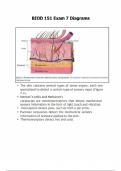
● The skin contains several types of sense organs, each one specialized to detect a certain type of sensory input (Figure 7.2). ● Merkel’s cells and Meissner's corpuscles are mechanoreceptors that detect mechanical sensory information in the form of light touch and vibration. ● Nociceptors detect pain, such as from a pin prick. ● Pacinian corpuscles detect the mechanical sensory information of pressure applied to the skin. ● Thermoreceptors detect hot and cold. ● Exocrine glands can be categorized by their shape and structure. ● Most glands in the body are multicellular glands, which are set within epithelial tissue and release their contents through a duct to the surface. ● Sebaceous glands (oil glands) produce sebum (Figure 7.3). ● Sebum helps to keep the skin and hair from drying out and inhibits the growth of harmful microorganisms. ● Sweat glands (sudoriferous glands) produce sweat in almost every part of the skin. The release of sweat is called perspiration. ● Perspiration helps the body to regulate its temperature through a process called thermoregulation by releasing heat from the body. Epidermis ● The epidermis (outermost layer of skin) is made up of stratified squamous epithelium. (Stratified means layered, and squamous means flat). ● The epidermis is avascular, meaning it does not have a direct blood supply. ● The epidermis receives its nutrients from the basement membrane (basal lamina). ● The epidermis is comprised of four distinct layers (listed from superficial to deep): the stratum corneum, stratum granulosum, stratum spinosum, and stratum basale (Figure 7.4). ● The stratum basale is firmly adhered to the basement membrane, which contains the blood supply for the epidermis. ● The cells of the stratum basale are constantly dividing to rebuild the skin. As cells continue to divide, they are pushed towards the external surface until they are dead and slough off (are removed through washing or friction). The most superficial layer of the epidermis is dead because it is far away from the blood and nutrient supply of the basement membrane. It takes about 30 days for a single cell to move from the stratum basale to the external surface of the stratum corneum. Dead cells remain within the stratum corneum for about another two weeks before they slough off. ● The stratum basale forms epidermal ridges (notice the grooves present in Figure 7.4 within the stratum basale). ● Epidermal ridges are unique to each person and consistent throughout a person’s lifetime. These projections extend all the way through the epidermis and form a distinct pattern commonly called fingerprints. Some of the cells found in the epidermis are keratinocytes, Langerhans cells, fibroblast cells, melanocytes, and Merkel cells (Figure 7.5). ● Melanocytes produce a pigment called melanin. ● Langerhans cells act as a first line of defense for the body, helping to identify microorganisms before they reach the bloodstream. ● Merkel cells are a type of sensory receptor that respond to light touch. ● Keratinocytes produce a tough substance called keratin. Cells within the epidermis undergo keratinization, which protects exposed surfaces of skin. ● Fibroblast cells produce collagen, which helps to adhere the cells of the epidermis to each other and give the skin elasticity. Dermis ● Sweat glands, oil glands, and sensory organs are also found within the dermis. ● The dermis contains the sensory organs of Meissner corpuscles and Pacinian corpuscles (as labeled in Figure 7.2). ● The dermis is comprised of a superficial papillary region and the deeper reticular region. Both layers of the dermis contain collagen fibers and elastic fibers, which enable the skin to be stretched (within certain limits) to prevent damage to underlying tissue. ● The dermis has a higher water content compared to the epidermis. A person who is dehydrated has decreased water content in the dermal layer of their skin. A person can be tested for dehydration by pinching the skin on the back of the hand. One indicator of dehydration is when the skin does not recoil back to its normal shape but instead stays pinched. ● The reticular region of the dermis contains the main blood supply for the skin. There is an extensive network of blood vessels to supply the skin with its needed nutrients. Branches come off the main arteries to supply nerves, glands, and hair follicles within the skin. ● A bruise, or contusion, is damage to one of these blood vessels. Blood leaks out of a blood vessel into the dermis, creating the color of the bruise (typically black and blue). Hypodermis ● a layer made mostly of fat cells (adipocytes). ● There are two layers: subcutaneous fat and subcutaneous tissue, which blend together. ● The hypodermis helps anchor the dermis to a layer of fat cells. ● Fat helps to insulate and regulate body temperature. ● The thickness of the hypodermis protects the deeper tissues
Document information
- Uploaded on
- April 30, 2025
- Number of pages
- 14
- Written in
- 2024/2025
- Type
- Exam (elaborations)
- Contains
- Questions & answers


