Dysrhythmia’s
Disorder of impulse formation, conduction of impulses, or both
4 properties
Automaticity – cells in SA node, atria, AV node, and bundle of His and purkinje fibers, which can
fire spontaneously
o Spontaneously fires 60-100 times/min
Excitability
Conductivity
Contractility
Diagnostics
ECG/EKG – electrical impulses produced in the heart
o Different wave forms to determine what is exactly wrong with the heart.
o 12 leads
Lead II is what we usually look at
o Shows changes suggesting structural changes, conduction disturbances, damage
(ischemia, infarction), lyte imbalances, or drug toxicity.
o Prep – will see artifact on monitor if leads are not secure
shave chest
Rub skin with dry gauze until slightly pink
Wipe with alcohol if oily skin
If diaphoretic apply skin protectant
o Calculate HR = count # of QRS complex in 1 min, # of R-R intervals in 6 seconds x 10
Telemetry – monitoring heart from a far
o R = clouds over grass (white and green)
o L = Smoke over fire (black and red)
o Middle = chocolate close to the heart (brown)
Assessment of heart rhythm
Make accurate interpretation and immediately assess clinical status of pt.
Are they hemodynamically stable?
Determine cause – priority!
o EX: fever tachy decreased CO and hypotension
Assess/treat pt., not monitor
D
D
D
D
D
D
P wave – Time of electrical impulse through atrium causing atrial depolarization; when atrial
contract
, o If missing/abnormal = issue with atrium
PR interval – time just before atrial contraction to just before ventricular contraction
QRS complex- electrical impulse running through ventricles ; when ventricles contract ]
o Normal time = 0.12 seconds
ST segment – Period where myocardium maintains contraction to expel blood from ventricles
o Should be flat
o Change = ischemia, injury, or infarction
o STEMI = infarction = coronary artery blocked and heart muscle can’t receive blood
o NSTEMI = less serious form of MI
T wave – Repolarization of ventricles
o Changes = hyper/hypokalemia, ischemia, or infarction
QT interval – Time electrical impulse first reaches ventricle through ventricular contraction (ORS)
to contraction (T)
o Shortens as HR increases
Teaching
Understand basic rhythm strips
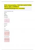
Sinus Bradycardia (<60 bpm) same rhythm. Here you see four R’s
o Symptomatic bradycardia = HR < 60 fatigue, dizziness, chest pain, syncope
o Associations
Regular during sleep or aerobically trained athletes
Sinus massage, Valsalva maneuver, hypothermia, increased IOP, vagal
stimulation, and certain drugs (B-blockers, CCB)
Diseases – hypothyroidism, increase ICP, and inferior MI
o Manifestations (if heart is NOT perfusing)
Pale, cool skin
Hypotension
Weakness
Angina
Dizziness/syncope
Confusion
SOB
o *If asymptomatic then they are perfusing- look in Giddins*
o Treatment
Atropine (anticholinergic)
Disorder of impulse formation, conduction of impulses, or both
4 properties
Automaticity – cells in SA node, atria, AV node, and bundle of His and purkinje fibers, which can
fire spontaneously
o Spontaneously fires 60-100 times/min
Excitability
Conductivity
Contractility
Diagnostics
ECG/EKG – electrical impulses produced in the heart
o Different wave forms to determine what is exactly wrong with the heart.
o 12 leads
Lead II is what we usually look at
o Shows changes suggesting structural changes, conduction disturbances, damage
(ischemia, infarction), lyte imbalances, or drug toxicity.
o Prep – will see artifact on monitor if leads are not secure
shave chest
Rub skin with dry gauze until slightly pink
Wipe with alcohol if oily skin
If diaphoretic apply skin protectant
o Calculate HR = count # of QRS complex in 1 min, # of R-R intervals in 6 seconds x 10
Telemetry – monitoring heart from a far
o R = clouds over grass (white and green)
o L = Smoke over fire (black and red)
o Middle = chocolate close to the heart (brown)
Assessment of heart rhythm
Make accurate interpretation and immediately assess clinical status of pt.
Are they hemodynamically stable?
Determine cause – priority!
o EX: fever tachy decreased CO and hypotension
Assess/treat pt., not monitor
D
D
D
D
D
D
P wave – Time of electrical impulse through atrium causing atrial depolarization; when atrial
contract
, o If missing/abnormal = issue with atrium
PR interval – time just before atrial contraction to just before ventricular contraction
QRS complex- electrical impulse running through ventricles ; when ventricles contract ]
o Normal time = 0.12 seconds
ST segment – Period where myocardium maintains contraction to expel blood from ventricles
o Should be flat
o Change = ischemia, injury, or infarction
o STEMI = infarction = coronary artery blocked and heart muscle can’t receive blood
o NSTEMI = less serious form of MI
T wave – Repolarization of ventricles
o Changes = hyper/hypokalemia, ischemia, or infarction
QT interval – Time electrical impulse first reaches ventricle through ventricular contraction (ORS)
to contraction (T)
o Shortens as HR increases
Teaching
Understand basic rhythm strips
Sinus Bradycardia (<60 bpm) same rhythm. Here you see four R’s
o Symptomatic bradycardia = HR < 60 fatigue, dizziness, chest pain, syncope
o Associations
Regular during sleep or aerobically trained athletes
Sinus massage, Valsalva maneuver, hypothermia, increased IOP, vagal
stimulation, and certain drugs (B-blockers, CCB)
Diseases – hypothyroidism, increase ICP, and inferior MI
o Manifestations (if heart is NOT perfusing)
Pale, cool skin
Hypotension
Weakness
Angina
Dizziness/syncope
Confusion
SOB
o *If asymptomatic then they are perfusing- look in Giddins*
o Treatment
Atropine (anticholinergic)