1. Overview of the Appendicular Skeleton
● Appendicular Skeleton: Composed of 126 bones (out of the total 206) that support the
limbs (arms and legs), including the pectoral (shoulder) and pelvic (hip) girdles.
● Function:
1. Supports and moves the axial skeleton.
2. Manipulates objects in the environment via the limbs.
● Major Regions:
1. Pectoral (shoulder) girdle
2. Arms and forearms
3. Wrists, hands, and fingers
4. Pelvic (hip) girdle
5. Thighs and lower legs
6. Ankles, feet, and toes
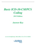
2. The Pectoral Girdle
● Bones: Clavicle (collarbone) and Scapula (shoulder blade).
● Location & Function:
○ Connects upper limbs to the axial skeleton.
○ Provides attachment sites for numerous skeletal muscles.
● Clavicle (Collarbone):
○ Shape varies by perspective: “S” shape from above (superior view), appears
straight from the front (anterior view).
○ Acts as a brace to keep the scapula in place.
○ Key parts:
■ Sternal (medial) end: articulates with the manubrium of the sternum.
■ Shaft (body).
■ Acromial (lateral) end: articulates with the acromion process of the
scapula.
● Scapula (Shoulder Blade):
○ Triangular, lies on the posterior/superior rib cage (ribs 2–7).
○ Borders/Edges: medial, lateral, superior.
○ Angles: superior, inferior, lateral.
○ Important Processes:
■ Spine of scapula → Acromion process (articulates with clavicle).
■ Coracoid process (muscle/ligament attachment; stabilizes shoulder
joint).
○ Glenoid cavity (fossa): articulates with the head of the humerus (shoulder joint).
○ Fossae for muscle attachments: supraspinous, infraspinous, subscapular.
,3. The Upper Limbs
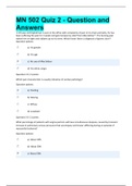
3.1 Humerus (Upper Arm Bone)
● Proximal Features:
○ Head: ball-shaped, fits into scapula’s glenoid cavity (forms the shoulder joint).
○ Anatomical neck: groove around the head.
○ Greater & Lesser tubercles: attachment sites for rotator cuff muscles.
○ Bicipital groove: between the tubercles for the biceps brachii tendon.
○ Surgical neck: common site of fractures.
● Mid-shaft:
○ Deltoid tuberosity: deltoid muscle attachment.
● Distal Features:
○ Capitulum (lateral): articulates with the radius.
○ Trochlea (medial): articulates with the ulna.
○ Lateral & Medial epicondyles: sites for forearm muscle attachments.
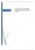
3.2 The Forearm (Radius & Ulna)
● General: Radius (lateral, thumb side) + Ulna (medial, pinky side). Connected by an
interosseous membrane.
● Ulna:
○ Proximal end:
■ Trochlear notch: articulates with humerus trochlea.
■ Olecranon process: bony prominence of the elbow (posterior).
■ Coronoid process: fits into humerus coronoid fossa (anterior).
■ Radial notch: articulates with radius head (proximal radioulnar joint).
○ Distal end:
■ Ulnar styloid process (small, medial wrist bump).
● Radius:
○ Proximal end:
■ Radial head: articulates with the capitulum (humerus) & radial notch
(ulna).
■ Radial neck → radial tuberosity: biceps brachii attachment.
○ Distal end:
■ Ulnar notch: articulates with the distal ulna (distal radioulnar joint).
■ Radial styloid process: forms lateral boundary of the wrist. (Radius is
wider distally.)
3.3 Wrist, Hand, and Fingers
, ● Carpal bones (Wrist): 8 short bones in two rows of four.
○ Proximal row (lateral to medial): Scaphoid, Lunate, Triquetrum, Pisiform.
○ Distal row (lateral to medial): Trapezium, Trapezoid, Capitate, Hamate.
● Hand (Metacarpals): 5 long bones numbered I–V (thumb to pinky). Each has a base
(proximal), shaft (middle), and head (distal = knuckles).
● Fingers (Phalanges): 14 total per hand.
○ Fingers II–V = 3 bones each (proximal, middle, distal).
○ Thumb (I) = 2 bones (proximal, distal).
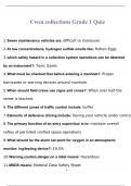
4. The Pelvic Girdle (Hip Bones)
● General:
○ Two coxal (hip) bones → each fuses from ilium, ischium, and pubis by ~age
13–15.
○ Articulates posteriorly with the sacrum (axial skeleton) at sacroiliac joints.
○ Right/left coxal bones meet anteriorly at the pubic symphysis.
○ Acetabulum: deep socket formed by fusion of ilium, ischium, and pubis,
articulates with the femur’s head (hip joint).
○ Obturator foramen: large opening formed by ischium and pubis for nerves and
blood vessels.
4.1 Ilium
● Largest of the hip bones, forms the superior portion.
● Key Landmarks:
○ Iliac crest: top ridge (the “hands on hips” area).
○ Anterior superior iliac spine (ASIS), anterior inferior iliac spine (AIIS),
posterior superior iliac spine (PSIS), and posterior inferior iliac spine (PIIS)
→ all are muscle attachment sites.
○ Greater sciatic notch: below the PIIS; passage for the sciatic nerve.
○ Iliac fossa (internal, concave surface).
4.2 Ischium
● Forms the posteroinferior (lower) part of the hip (your “sit bones”).
● Ischial tuberosities: roughened area that bears body weight when seated; also muscle
attachments.
4.3 Pubis
● Most anterior (front) and inferior region of the hip bone.
● Pubic symphysis: fibrocartilage pad where left and right pubic bones meet anteriorly.
● Appendicular Skeleton: Composed of 126 bones (out of the total 206) that support the
limbs (arms and legs), including the pectoral (shoulder) and pelvic (hip) girdles.
● Function:
1. Supports and moves the axial skeleton.
2. Manipulates objects in the environment via the limbs.
● Major Regions:
1. Pectoral (shoulder) girdle
2. Arms and forearms
3. Wrists, hands, and fingers
4. Pelvic (hip) girdle
5. Thighs and lower legs
6. Ankles, feet, and toes
2. The Pectoral Girdle
● Bones: Clavicle (collarbone) and Scapula (shoulder blade).
● Location & Function:
○ Connects upper limbs to the axial skeleton.
○ Provides attachment sites for numerous skeletal muscles.
● Clavicle (Collarbone):
○ Shape varies by perspective: “S” shape from above (superior view), appears
straight from the front (anterior view).
○ Acts as a brace to keep the scapula in place.
○ Key parts:
■ Sternal (medial) end: articulates with the manubrium of the sternum.
■ Shaft (body).
■ Acromial (lateral) end: articulates with the acromion process of the
scapula.
● Scapula (Shoulder Blade):
○ Triangular, lies on the posterior/superior rib cage (ribs 2–7).
○ Borders/Edges: medial, lateral, superior.
○ Angles: superior, inferior, lateral.
○ Important Processes:
■ Spine of scapula → Acromion process (articulates with clavicle).
■ Coracoid process (muscle/ligament attachment; stabilizes shoulder
joint).
○ Glenoid cavity (fossa): articulates with the head of the humerus (shoulder joint).
○ Fossae for muscle attachments: supraspinous, infraspinous, subscapular.
,3. The Upper Limbs
3.1 Humerus (Upper Arm Bone)
● Proximal Features:
○ Head: ball-shaped, fits into scapula’s glenoid cavity (forms the shoulder joint).
○ Anatomical neck: groove around the head.
○ Greater & Lesser tubercles: attachment sites for rotator cuff muscles.
○ Bicipital groove: between the tubercles for the biceps brachii tendon.
○ Surgical neck: common site of fractures.
● Mid-shaft:
○ Deltoid tuberosity: deltoid muscle attachment.
● Distal Features:
○ Capitulum (lateral): articulates with the radius.
○ Trochlea (medial): articulates with the ulna.
○ Lateral & Medial epicondyles: sites for forearm muscle attachments.
3.2 The Forearm (Radius & Ulna)
● General: Radius (lateral, thumb side) + Ulna (medial, pinky side). Connected by an
interosseous membrane.
● Ulna:
○ Proximal end:
■ Trochlear notch: articulates with humerus trochlea.
■ Olecranon process: bony prominence of the elbow (posterior).
■ Coronoid process: fits into humerus coronoid fossa (anterior).
■ Radial notch: articulates with radius head (proximal radioulnar joint).
○ Distal end:
■ Ulnar styloid process (small, medial wrist bump).
● Radius:
○ Proximal end:
■ Radial head: articulates with the capitulum (humerus) & radial notch
(ulna).
■ Radial neck → radial tuberosity: biceps brachii attachment.
○ Distal end:
■ Ulnar notch: articulates with the distal ulna (distal radioulnar joint).
■ Radial styloid process: forms lateral boundary of the wrist. (Radius is
wider distally.)
3.3 Wrist, Hand, and Fingers
, ● Carpal bones (Wrist): 8 short bones in two rows of four.
○ Proximal row (lateral to medial): Scaphoid, Lunate, Triquetrum, Pisiform.
○ Distal row (lateral to medial): Trapezium, Trapezoid, Capitate, Hamate.
● Hand (Metacarpals): 5 long bones numbered I–V (thumb to pinky). Each has a base
(proximal), shaft (middle), and head (distal = knuckles).
● Fingers (Phalanges): 14 total per hand.
○ Fingers II–V = 3 bones each (proximal, middle, distal).
○ Thumb (I) = 2 bones (proximal, distal).
4. The Pelvic Girdle (Hip Bones)
● General:
○ Two coxal (hip) bones → each fuses from ilium, ischium, and pubis by ~age
13–15.
○ Articulates posteriorly with the sacrum (axial skeleton) at sacroiliac joints.
○ Right/left coxal bones meet anteriorly at the pubic symphysis.
○ Acetabulum: deep socket formed by fusion of ilium, ischium, and pubis,
articulates with the femur’s head (hip joint).
○ Obturator foramen: large opening formed by ischium and pubis for nerves and
blood vessels.
4.1 Ilium
● Largest of the hip bones, forms the superior portion.
● Key Landmarks:
○ Iliac crest: top ridge (the “hands on hips” area).
○ Anterior superior iliac spine (ASIS), anterior inferior iliac spine (AIIS),
posterior superior iliac spine (PSIS), and posterior inferior iliac spine (PIIS)
→ all are muscle attachment sites.
○ Greater sciatic notch: below the PIIS; passage for the sciatic nerve.
○ Iliac fossa (internal, concave surface).
4.2 Ischium
● Forms the posteroinferior (lower) part of the hip (your “sit bones”).
● Ischial tuberosities: roughened area that bears body weight when seated; also muscle
attachments.
4.3 Pubis
● Most anterior (front) and inferior region of the hip bone.
● Pubic symphysis: fibrocartilage pad where left and right pubic bones meet anteriorly.