Combine Practical Exam and Final Exam- Microbiology Review sheet
Practical Exam
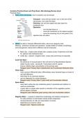
● Parts of the microscope, how to properly use microscope
Transport - carry with two hands- one on the arm of the
microscope, one under the base
Cleaning: use only lens paper and also clean the
condenser lens
Operation:
- turn the light lamp on
- move the condenser to the highest position
- move the scanning (4x) or low power (10x)
objective lens into place
● Stains: be able to interpret differential stains, also know reagents used
Staining : enhances contrast and resolution: reveals details of cellular morphology
and arrangement, stains bind to different cell and molecules
❖ Basic dye - crystal violet (simple stain, prepare smear of specimen and heat
fix, safranin and methylene blue)
❖ Acidic dye - nigrosin ( negative stain, smear prep and fixing not required)
Acid Fast Stain :
- the presence of unusual lipids in the cell wall of my Mycobacterial Species
called mycolic acids , causing M. Tuberculosis and M. Leprae
- Kinyoun Method high concentration of the primary stain
Reagent used: Primary Stain Carbolfuchsin and Counterstain Methylene Blue
Gram Stain :
- most common differential stain; differentiates many bacterial types on basis of
cell wall difference
- initial step for an unknown bacteria
- can be diagnostic for certain diseases
Reagent used: Primary Stain Crystal violet and Counterstain Safranin
Spore Stain:
- uses heat to allow the dye (malachite green) to penetrate and (safranin is
used as counterstain
- a gram stain or simple stain results in coloration of the vegetative portion,
spores appear colorless
Reagents used: Malachite green primary stain and counterstain safranin
Negative Stain : - purpose to determine morphology and cellular arrangement
- for cells sensitive to heat fixation during smear preparation
- minimal distortion of cells (no heat fixation involved)
- staphylococcus aureus culture
Reagent used : Nigrosin acidic dye
, ● Streak Plate Method: - divide the plate into 4 quadrants using a wire loop
- hold the loop flat against the agar and streak across surface
- reflame the loop before changing direction of streaking
The inoculum source can be from
- liquid culture
- plate culture
- slant
● Aseptic Technique - to minimize contamination
● Differential and Selective Media
● Biochemical Tests (Lab Final Portion lab 7-11)
● Knowledge of proper biosafety procedures
-One should wash hands before lab starts and after lab ends
-This is a BSL 2 lab, bacterial strains designated as BSL-1 or BSL-2 can be used in
here
-There are occasions when it is proper procedure to discard paper towels in
biohazard waste
Practical Exam
● Parts of the microscope, how to properly use microscope
Transport - carry with two hands- one on the arm of the
microscope, one under the base
Cleaning: use only lens paper and also clean the
condenser lens
Operation:
- turn the light lamp on
- move the condenser to the highest position
- move the scanning (4x) or low power (10x)
objective lens into place
● Stains: be able to interpret differential stains, also know reagents used
Staining : enhances contrast and resolution: reveals details of cellular morphology
and arrangement, stains bind to different cell and molecules
❖ Basic dye - crystal violet (simple stain, prepare smear of specimen and heat
fix, safranin and methylene blue)
❖ Acidic dye - nigrosin ( negative stain, smear prep and fixing not required)
Acid Fast Stain :
- the presence of unusual lipids in the cell wall of my Mycobacterial Species
called mycolic acids , causing M. Tuberculosis and M. Leprae
- Kinyoun Method high concentration of the primary stain
Reagent used: Primary Stain Carbolfuchsin and Counterstain Methylene Blue
Gram Stain :
- most common differential stain; differentiates many bacterial types on basis of
cell wall difference
- initial step for an unknown bacteria
- can be diagnostic for certain diseases
Reagent used: Primary Stain Crystal violet and Counterstain Safranin
Spore Stain:
- uses heat to allow the dye (malachite green) to penetrate and (safranin is
used as counterstain
- a gram stain or simple stain results in coloration of the vegetative portion,
spores appear colorless
Reagents used: Malachite green primary stain and counterstain safranin
Negative Stain : - purpose to determine morphology and cellular arrangement
- for cells sensitive to heat fixation during smear preparation
- minimal distortion of cells (no heat fixation involved)
- staphylococcus aureus culture
Reagent used : Nigrosin acidic dye
, ● Streak Plate Method: - divide the plate into 4 quadrants using a wire loop
- hold the loop flat against the agar and streak across surface
- reflame the loop before changing direction of streaking
The inoculum source can be from
- liquid culture
- plate culture
- slant
● Aseptic Technique - to minimize contamination
● Differential and Selective Media
● Biochemical Tests (Lab Final Portion lab 7-11)
● Knowledge of proper biosafety procedures
-One should wash hands before lab starts and after lab ends
-This is a BSL 2 lab, bacterial strains designated as BSL-1 or BSL-2 can be used in
here
-There are occasions when it is proper procedure to discard paper towels in
biohazard waste