🔬
Histologie
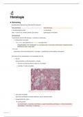
Beenmerg
hematopoeise, fagocytering, maturatie B-lymfocyten
Rood beenmerg Geel beenmerg
= hematopoietisch actief ⇒ vetweefsel
alles → wervel, rib, schedel, bekken, prox femur (pqthologisch reversibel)
Hematopoeise:
1 pluripotente stamcel (IM kleuring) → meerdere in beenmerg
→ multipotente stamcellen:
lymfoïde voorlopercellen en myeloïde voorlopercellen
respectievelijk B- en T-lymfocyten, en de granulocyten, monocyten, erythrocyten, megakaryocyten,
dentritische cellen, mast cellen
G-CSF
granulocyt colony stimulating factor / neurogen → groeifactor als behandeling neutropenie
beenmerg zit tsss bottrabekels en het medullaire bot met
stroma
reticulumcellen en reticulinevezels = netwerk
ertussen de hemtopoeitische cellen en macrofagen
vetcellen // locatie en leeftijd
vasculaire sinusoïden
discontinu endotheel en basale membraan, weg nr bloedcirculatie
hematopoietische cellen
erythropoiese
pro-erythroblast → basofiele eryhtroblast → polychromatofiele erythroblast → orthochromatofiele
erythroblast → reticulocyt → erythrocyt
Histologie 1
, → celvolume daalt, nr pyknotische kern en zelfs uitgeworpen
afname polyribosomen (basofiel) maar toename Hg (eosinofiel)
→ vormen erythronen met gespecialiseerde macrofagen als Fe-aanvoer
Erythropoietine > nier
onderscheiding via GIemsa-kleuring
granulopoiese
myeloblast (grote kern, fijne chromatine, 1-3 nucleolen en basofiel)
promyelocyt (grootste, azurofiele granulen met lysosomale EZ en myeoloperoxidase)
3 types myelocyten: (herkenbare spec granules, bij maturatie meer granules, condensere kern en
afname kernvolume)
neutrofiele myelocyt eosinofiele myelocyt basofiele myelocyt
neutrofiele metamyelocyt eosinofiele metamyelocyt basofiele metamyelocyt
staaf eosinofiele granulocyten basofiele granulocyten
neutrofiele granulocyte
* De ontw v/d mature
neutrofiele granulocyten
vindt plaats in 4
compartimenten:
granulopoietisch
compartiment in actief
beenmerg
stapeling van mature
cellen in beenmerg
circulerende populatie
marginerende populatie
dat bindt op endotheel om
transiënt te accumuleren
en zo bij inspanning de
circulerende te vervoegen
Ook via Giemsa-kleuring te onderscheiden
maturatie monocyten
monoblast = committed progenitor cel // myeloblast
promonocyt (kernplooitje)
monocyt (ruwer ER, cytoplasme, etc)
→ verlaat beenmerg onmiddelijk → uren/dagen in bloed → weefsels uitrijpen tot macrofagen
(overleven tot maanden)
maturatie lymfocyten
→ thymus → T-lymfocyten → secundaire lymfoïde organen (lymfoblasten licht optisch herkenbaar)
→ B-lymfocyten (lymfoblasten gering en moeilijk zichtbaar)
vorming bloedplaatjes
megakaryoblasten
endomitosis (DNA-replicatie zonder celdeling) → polyplo¨die (8-64N)
megakyrocyten (tot 150 μm diameter, onregelamtig gelobde kern, polyploïd DNA, sterk ER, groot
Golgi (→ granulen bloedplaatjes)) geven de bleodplaatjes rehctstreeks af in de sinusoïden via
Histologie 2
, proplatelets (diens fragmentatie)
> thrombopoietine
histo plaatjes:
rode bloedlichaampjes: rode, ronde cellen zonder kern maar met een centrale indeuking
witte bloedcellen:
neutrofielen (meest voorkomend, sterk gesegmenteerde kern)
eosinofielen (oranje, korrelig cytoplasma en brilvormige kern)
lymfocyten (grote, donkere kern en een geringe hoeveelheid cytoplasma)
monocyten (grote cellen met een hoefijzervormige tot niervormige kern)
basofielen (blauw tot violet gekleurde korrels in het cytoplasma)
bloedplaatjes: kleine, donker gekleurde, schijfvormige fragmenten (kernloos)
Histologie 3
Histologie
Beenmerg
hematopoeise, fagocytering, maturatie B-lymfocyten
Rood beenmerg Geel beenmerg
= hematopoietisch actief ⇒ vetweefsel
alles → wervel, rib, schedel, bekken, prox femur (pqthologisch reversibel)
Hematopoeise:
1 pluripotente stamcel (IM kleuring) → meerdere in beenmerg
→ multipotente stamcellen:
lymfoïde voorlopercellen en myeloïde voorlopercellen
respectievelijk B- en T-lymfocyten, en de granulocyten, monocyten, erythrocyten, megakaryocyten,
dentritische cellen, mast cellen
G-CSF
granulocyt colony stimulating factor / neurogen → groeifactor als behandeling neutropenie
beenmerg zit tsss bottrabekels en het medullaire bot met
stroma
reticulumcellen en reticulinevezels = netwerk
ertussen de hemtopoeitische cellen en macrofagen
vetcellen // locatie en leeftijd
vasculaire sinusoïden
discontinu endotheel en basale membraan, weg nr bloedcirculatie
hematopoietische cellen
erythropoiese
pro-erythroblast → basofiele eryhtroblast → polychromatofiele erythroblast → orthochromatofiele
erythroblast → reticulocyt → erythrocyt
Histologie 1
, → celvolume daalt, nr pyknotische kern en zelfs uitgeworpen
afname polyribosomen (basofiel) maar toename Hg (eosinofiel)
→ vormen erythronen met gespecialiseerde macrofagen als Fe-aanvoer
Erythropoietine > nier
onderscheiding via GIemsa-kleuring
granulopoiese
myeloblast (grote kern, fijne chromatine, 1-3 nucleolen en basofiel)
promyelocyt (grootste, azurofiele granulen met lysosomale EZ en myeoloperoxidase)
3 types myelocyten: (herkenbare spec granules, bij maturatie meer granules, condensere kern en
afname kernvolume)
neutrofiele myelocyt eosinofiele myelocyt basofiele myelocyt
neutrofiele metamyelocyt eosinofiele metamyelocyt basofiele metamyelocyt
staaf eosinofiele granulocyten basofiele granulocyten
neutrofiele granulocyte
* De ontw v/d mature
neutrofiele granulocyten
vindt plaats in 4
compartimenten:
granulopoietisch
compartiment in actief
beenmerg
stapeling van mature
cellen in beenmerg
circulerende populatie
marginerende populatie
dat bindt op endotheel om
transiënt te accumuleren
en zo bij inspanning de
circulerende te vervoegen
Ook via Giemsa-kleuring te onderscheiden
maturatie monocyten
monoblast = committed progenitor cel // myeloblast
promonocyt (kernplooitje)
monocyt (ruwer ER, cytoplasme, etc)
→ verlaat beenmerg onmiddelijk → uren/dagen in bloed → weefsels uitrijpen tot macrofagen
(overleven tot maanden)
maturatie lymfocyten
→ thymus → T-lymfocyten → secundaire lymfoïde organen (lymfoblasten licht optisch herkenbaar)
→ B-lymfocyten (lymfoblasten gering en moeilijk zichtbaar)
vorming bloedplaatjes
megakaryoblasten
endomitosis (DNA-replicatie zonder celdeling) → polyplo¨die (8-64N)
megakyrocyten (tot 150 μm diameter, onregelamtig gelobde kern, polyploïd DNA, sterk ER, groot
Golgi (→ granulen bloedplaatjes)) geven de bleodplaatjes rehctstreeks af in de sinusoïden via
Histologie 2
, proplatelets (diens fragmentatie)
> thrombopoietine
histo plaatjes:
rode bloedlichaampjes: rode, ronde cellen zonder kern maar met een centrale indeuking
witte bloedcellen:
neutrofielen (meest voorkomend, sterk gesegmenteerde kern)
eosinofielen (oranje, korrelig cytoplasma en brilvormige kern)
lymfocyten (grote, donkere kern en een geringe hoeveelheid cytoplasma)
monocyten (grote cellen met een hoefijzervormige tot niervormige kern)
basofielen (blauw tot violet gekleurde korrels in het cytoplasma)
bloedplaatjes: kleine, donker gekleurde, schijfvormige fragmenten (kernloos)
Histologie 3