Lecture 1: Tissue segmentation and structural statistics
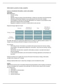
Tissue-type segmentation
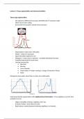
- We segment 3 diffferent tissue types (GM,WM,CSF) intensity model
- Other way of color coding
- Every peak in the graph is another tissue structure
- Segmentation made easier with peaks
- Model = mixture of Gaussians
- Signal intensities depicted in red
- Histogram = voxel count vs intensity = probably distribution function
- Probably determined for each tissue
- Overlap worsened by
o Bias field
o Blurring
o Low resolution
o Head motion stripes coming in, change of intensity of tissue
o Noise
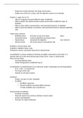
Histograms show peaks, when there is a bias, less visible peaks
Improving intensity segmentation with neighbourhood information = if my neighbours are GM, then
probably I am too.
- Higher probability of being a neighbour then not.
- K-means clusters, more robust to noise.
- Right balance needed between believing neighbours or intensity
, More neighbourhood information, the smaller CSF parts in grey become
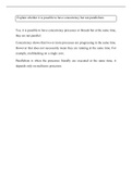
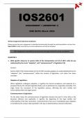
The intensity that is meeting the most, takes it all. Voxel can only be one of the 3. In reality this voxel
is only a very small mm part. This cube in space does not only need to have white or gray matter. If I
am imaging, a voxel that is on the border, each voxel should be allowed to be more than just 1. Each
voxel probability map. Light colours is very big probability of grey matter, darker is lower, by this
map you will get an image.
- 1st is voxel for PVE voxel for CSF
- 2nd is for grey matter
- 3rd is for white matter
- Grey-Ish colours is unsure, could be both white and grey matter structures, probability of
intensities
1. So first an approximate segmentation with the intensity model into CSF, WM, GM
2. Iterate estimate bias field
3. Estimate segmentation by looking at intensity and its neighbours
4. Apply partial volume model
5. MRF of mixel type, how many tissues
6. Partial volume estimation (PVE)
Besides of intensity model and PVE use of priors
- Average of previous subjects segmentations that have been researched before =
segmentation priors
- This is great alternative in case of bias or radio frequency disruption or strong motion
- Priors are called priors because = prior information is used; info known before the start
Tissue-type segmentation
- We segment 3 diffferent tissue types (GM,WM,CSF) intensity model
- Other way of color coding
- Every peak in the graph is another tissue structure
- Segmentation made easier with peaks
- Model = mixture of Gaussians
- Signal intensities depicted in red
- Histogram = voxel count vs intensity = probably distribution function
- Probably determined for each tissue
- Overlap worsened by
o Bias field
o Blurring
o Low resolution
o Head motion stripes coming in, change of intensity of tissue
o Noise
Histograms show peaks, when there is a bias, less visible peaks
Improving intensity segmentation with neighbourhood information = if my neighbours are GM, then
probably I am too.
- Higher probability of being a neighbour then not.
- K-means clusters, more robust to noise.
- Right balance needed between believing neighbours or intensity
, More neighbourhood information, the smaller CSF parts in grey become
The intensity that is meeting the most, takes it all. Voxel can only be one of the 3. In reality this voxel
is only a very small mm part. This cube in space does not only need to have white or gray matter. If I
am imaging, a voxel that is on the border, each voxel should be allowed to be more than just 1. Each
voxel probability map. Light colours is very big probability of grey matter, darker is lower, by this
map you will get an image.
- 1st is voxel for PVE voxel for CSF
- 2nd is for grey matter
- 3rd is for white matter
- Grey-Ish colours is unsure, could be both white and grey matter structures, probability of
intensities
1. So first an approximate segmentation with the intensity model into CSF, WM, GM
2. Iterate estimate bias field
3. Estimate segmentation by looking at intensity and its neighbours
4. Apply partial volume model
5. MRF of mixel type, how many tissues
6. Partial volume estimation (PVE)
Besides of intensity model and PVE use of priors
- Average of previous subjects segmentations that have been researched before =
segmentation priors
- This is great alternative in case of bias or radio frequency disruption or strong motion
- Priors are called priors because = prior information is used; info known before the start