Lecture 1:
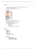
- 100 billion neurons in the brain, each connected to 1000-100.000 other neurons.
- Around 15000 synapses per neuron
- Different types of neurons have different functions
- The dendrites receive info from other neurons
- The axons conduct information via action potentials
- Glial cells support the neurons
- Structural, metabolic and functional support
o Nutrient transport
o NGF repair
o Isolation axons
o Controls composition extracellular fluid
Anatomy
- Dendrites
- Nucleus
- Myelin sheaths
- Axon
- Synaptic cleft
- Vesicles neurotransmitters
- Neuroglia
o Microglia = defense
o Astrocyte = support/nutrition
o Oligodendrocytes = insulation
,Axon is surrounded by major and minor dense lines with spaces in between called the paranodal
pockets. Oligodendrocytes are connected to the axons by pedicles. The outer layer of the axon Is
called the cytoplasmic flange
- CNS
o Brain → gyri and sulci, sulci are the folds
o Spinal cord
- PNS
o Cranial nerve
o Spinal nerve
- Brain is protected by skull
- CNS in skull and vertebral column
- Functions:
o Perception, movement, learning memory, personality, emotions behaviour,
consciousness, language
- Most of neurons are interneurons
,- Afferent/ sensory input → interneurons → efferent/motor output
- Neo cortex contains of 6 neuron layers
- Light part is white matter
o Fibre tracts
- Dark is grey matter/ cortex/ nuclei
o Nucleus = group of cell bodies
- Basal ganglia → initiation and organization of motor movements
Of the butterfly → upper is posterior, middle is lateral, lower is anterior
• Above arrow is spinal cord with grey matter = canalis centralis
• Lower arrow is white matter, located peripherally and contains ascending and
descending fibre tracts
, - Embryological development → neural plate fold tube from the surface = ectoderm
- Eventually neural tube closes → when it goes wrong with closing a spina bifida can happen
- On the side of the neural tube cavity, the neuroepithelial cells move and change in the
different cells (neuroblast, neurons, glioblast, radial glia, ependymal cell via mitosis
From marginal layer, to mantle zone to ependymal zone/ ventricular layer