Bontrager Chapter 2 Workbook Guaranteed Success
Summary of Thoracic Anatomy and Radiographic Principles
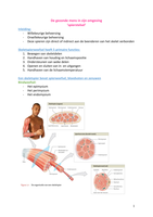
1. Components of the Bony Thorax:
- (A) Sternum
- (B) Clavicles
- (C) Scapulae
- (D) Ribs
- (E) Thoracic vertebrae
2. Landmarks for Central Ray Location:
- (A) Vertebra prominens (spinous process on the seventh cervical vertebra, C7)
- (B) Jugular notch (upper portion of the sternum)
3. Divisions of the Respiratory System:
✔️ Pharynx, Trachea, Bronchi, Lungs
4. Anatomical Terms:
- Adam's apple: ✔️ Thyroid cartilage
- Voice box: ✔️ Larynx
- Breastbone: ✔️ Sternum
- Shoulder: ✔️ Scapula
- Collarbone: ✔️ Clavicle
5. Divisions of the Pharynx:
✔️ 1. Nasopharynx
✔️ 2. Oropharynx
✔️ 3. Laryngopharynx
, 6. Structure Preventing Entry of Food into the Respiratory System:
✔️ Epiglottis
7. Trachea and Esophagus Relationship:
✔️ The trachea is located anteriorly to the esophagus.
8. Anatomical Structure in Anterior Neck:
✔️ Hyoid bone
9. Bronchus Particle Inhalation:
- A. Right bronchus
- B. Why? It is larger in diameter and more vertical.
10. Prominence Seen in Bronchi Division:
- A. Carina
- B. This prominence is at the level of the T5 vertebra.
11. Small Air Sacs for Gas Exchange:
✔️ Alveoli
12. Double-Walled Sac Containing the Lungs:
- A. Pleura
- B. Parietal pleura (outer layer)
- C. Inner layer adhering to the pulmonary or visceral pleura
- D. Pleural cavity (potential space between the pleura layers)
- E. Air in the pleural cavity results in pneumothorax.
Summary of Thoracic Anatomy and Radiographic Principles
1. Components of the Bony Thorax:
- (A) Sternum
- (B) Clavicles
- (C) Scapulae
- (D) Ribs
- (E) Thoracic vertebrae
2. Landmarks for Central Ray Location:
- (A) Vertebra prominens (spinous process on the seventh cervical vertebra, C7)
- (B) Jugular notch (upper portion of the sternum)
3. Divisions of the Respiratory System:
✔️ Pharynx, Trachea, Bronchi, Lungs
4. Anatomical Terms:
- Adam's apple: ✔️ Thyroid cartilage
- Voice box: ✔️ Larynx
- Breastbone: ✔️ Sternum
- Shoulder: ✔️ Scapula
- Collarbone: ✔️ Clavicle
5. Divisions of the Pharynx:
✔️ 1. Nasopharynx
✔️ 2. Oropharynx
✔️ 3. Laryngopharynx
, 6. Structure Preventing Entry of Food into the Respiratory System:
✔️ Epiglottis
7. Trachea and Esophagus Relationship:
✔️ The trachea is located anteriorly to the esophagus.
8. Anatomical Structure in Anterior Neck:
✔️ Hyoid bone
9. Bronchus Particle Inhalation:
- A. Right bronchus
- B. Why? It is larger in diameter and more vertical.
10. Prominence Seen in Bronchi Division:
- A. Carina
- B. This prominence is at the level of the T5 vertebra.
11. Small Air Sacs for Gas Exchange:
✔️ Alveoli
12. Double-Walled Sac Containing the Lungs:
- A. Pleura
- B. Parietal pleura (outer layer)
- C. Inner layer adhering to the pulmonary or visceral pleura
- D. Pleural cavity (potential space between the pleura layers)
- E. Air in the pleural cavity results in pneumothorax.