PULMONOLOGY USMLE STEP 1 NOTES
EMBRYOLOGY
The Lung bud which is also called the respiratory diverticulum is the outgrowth of the
foregut. This forms during the 4th week of development.
Lung Maturation Stages/Periods
Pseudoglandular (5-16 weeks)
Canalicular (16-26 weeks)
Saccular (26 weeks to birth)
Alveolar (after birth)
Anatomy
Bronchi → Hyaline cartilage
Bronchioles → No cartilage. Terminal → respiratory
Alveoli → Capillaries and gas exchange.
Pseudoglandular Period
In this period the lung resembles a gland. There is branching to level of terminal
bronchioles. No respiratory bronchioles or alveoli are present.
Fetal Respiration
Fetal breathing movements occur in utero. The baby aspirates amniotic fluid which
stimulates lung development and growth of respiratory muscles. Fetal respiration is
important for growth during pseudoglandular phase.
Canalicular Period
In this period the terminal bronchioles divide and form respiratory bronchioles.
Respiratory bronchioles divide into alveolar ducts. Survival after birth is possible at the
end of this period. The airway lumens become larger.
Type II pneumocytes form in this period. This pneumocyte produces surfactant to lower
surface tension and keeps alveoli open.
Saccular Period
Terminal sacs (primitive alveoli) form. Capillaries multiply in contact with alveoli.
Alveolar Period
At birth, only about ⅓ of alveoli is present. Following birth there is increasing number of
respiratory bronchioles and alveoli. There is continued lung development through age
10. Alveolarization: Airspaces are subdivided and new walls will be formed (septa)
,Bronchopulmonary Dysplasia
Occurs in premature infants and is treated in the NICU. Surfactants, oxygen,
mechanical ventilation. Oxygen toxicity and lung trauma. Alveolarization does not
progress normally. Respiratory problems during infancy. This often improves during
childhood.
Pulmonary Hypoplasia
This is seen in:
Oligohydramnios (Potter’s sequence)
Congenital diaphragmatic hernia
- This is defective formation of pleuroperitoneal membrane
- This leads to a hole in the diaphragm and abdominal organs herniate into chest.
If there is herniation in utero it will lead to pulmonary hypoplasia. Often fatal.
Bronchogenic Cysts
Abnormal budding of foregut. Usually found in the mediastinum. Contain clear fluid but
air is seen when it is infected. Bronchogenic cysts do not communicate with lungs.
Lined by respiratory epithelium (columnar, ciliated). The walls contain cartilage which is
a diagnostic criteria. Often asymptomatic. May lead to pneumonia, compression of
airway.
Pulmonary Vascular Resistance
In utero
- PVR is high
- Canalicular stage: few/no pulmonary capillaries
- Later stages: hypoxemia → vasoconstriction
- Umbilical venous blood: PaO2 30 mmHg; O2 saturation = 80%
- Only about 10% of cardiac output to lungs
At birth
- PVR falls significantly
- 100% cardiac output through lungs
ANATOMY
Zones
Conducting Zone
- No gas exchange
- Large airways: nose, pharynx, trachea and bronchi
- Filters, warms and humidifies air
Respiratory Zone
- Gas exchange
- Respiratory bronchioles, alveolar ducts and alveoli
,Mucous
Secretions produced by respiratory tracts. The secretions are mostly glycoproteins and
water. The mucus is secreted by goblet cells in bronchial walls. Protects against
particulates, infection. Beating cilia move mucus to epiglottis → swallowed
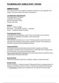
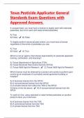
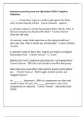
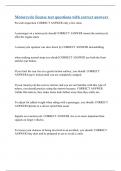
Alveoli
Alveoli are small sacs that is
responsible for gas exchange.
It is surrounded by capillaries.
Alveolar Cells: Pneumocytes
Type 1
- Most common (97% of cells)
- Thin for gas exchange
Type 2
- Produce surfactant
- Can proliferate to other cells - key for regeneration after injury
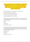
Club Cells (bronchioles)
- Surfactant
- Responsible for detoxification
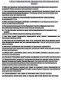
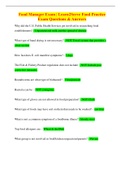
Surfactant
When you exhale the alveoli shrinks. The alveoli can collapse and this is called
atelectasis due to decreased efficiency gas exchange.
Surfactant allows alveoli to avoid collapse.
, Surfactants are secreted by type 2 pneumocytes. Mix of lecithins. Especially
dipalmitoylphosphatidylcholine
Fetal Lung Maturity
Lungs are “mature” when enough surfactant are present. Occurs around 35 weeks.
Lecithin-sphingomyelin ratio (L/S ratio)
Both produced equally until approx 35 weeks.
Ratio >2.0 in amniotic fluid suggests lungs mature
Preterm delivery: betamethasone is used to stimulate surfactant production in lungs.
Neonatal Respiratory Distress Syndrome
Hyaline membrane disease. Can lead to atelectasis. There is severe hypoxemia/↑pCO2
(poor ventilation). Poorly responsive to O2 because lungs are collapsed (alveoli) and
intrapulmonary shunting.
Risk Factors
- Prematurity
- Maternal diabetes: high insulin levels decrease surfactant production
- Cesarean delivery: lack of vaginal compression stress leads to reduced fetal
cortisol and reduction in surfactant
There are many complications in NRDS:
1. Bronchopulmonary dysplasia
2. Patent ductus arteriosus (hypoxia keeps shunt open)
3. Retinopathy of prematurity
- Oxygen → free radical formation
- Neovascularization in the retina
- Retinal detachment → blindness
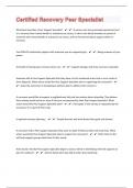
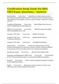
Lobes of the Lung
EMBRYOLOGY
The Lung bud which is also called the respiratory diverticulum is the outgrowth of the
foregut. This forms during the 4th week of development.
Lung Maturation Stages/Periods
Pseudoglandular (5-16 weeks)
Canalicular (16-26 weeks)
Saccular (26 weeks to birth)
Alveolar (after birth)
Anatomy
Bronchi → Hyaline cartilage
Bronchioles → No cartilage. Terminal → respiratory
Alveoli → Capillaries and gas exchange.
Pseudoglandular Period
In this period the lung resembles a gland. There is branching to level of terminal
bronchioles. No respiratory bronchioles or alveoli are present.
Fetal Respiration
Fetal breathing movements occur in utero. The baby aspirates amniotic fluid which
stimulates lung development and growth of respiratory muscles. Fetal respiration is
important for growth during pseudoglandular phase.
Canalicular Period
In this period the terminal bronchioles divide and form respiratory bronchioles.
Respiratory bronchioles divide into alveolar ducts. Survival after birth is possible at the
end of this period. The airway lumens become larger.
Type II pneumocytes form in this period. This pneumocyte produces surfactant to lower
surface tension and keeps alveoli open.
Saccular Period
Terminal sacs (primitive alveoli) form. Capillaries multiply in contact with alveoli.
Alveolar Period
At birth, only about ⅓ of alveoli is present. Following birth there is increasing number of
respiratory bronchioles and alveoli. There is continued lung development through age
10. Alveolarization: Airspaces are subdivided and new walls will be formed (septa)
,Bronchopulmonary Dysplasia
Occurs in premature infants and is treated in the NICU. Surfactants, oxygen,
mechanical ventilation. Oxygen toxicity and lung trauma. Alveolarization does not
progress normally. Respiratory problems during infancy. This often improves during
childhood.
Pulmonary Hypoplasia
This is seen in:
Oligohydramnios (Potter’s sequence)
Congenital diaphragmatic hernia
- This is defective formation of pleuroperitoneal membrane
- This leads to a hole in the diaphragm and abdominal organs herniate into chest.
If there is herniation in utero it will lead to pulmonary hypoplasia. Often fatal.
Bronchogenic Cysts
Abnormal budding of foregut. Usually found in the mediastinum. Contain clear fluid but
air is seen when it is infected. Bronchogenic cysts do not communicate with lungs.
Lined by respiratory epithelium (columnar, ciliated). The walls contain cartilage which is
a diagnostic criteria. Often asymptomatic. May lead to pneumonia, compression of
airway.
Pulmonary Vascular Resistance
In utero
- PVR is high
- Canalicular stage: few/no pulmonary capillaries
- Later stages: hypoxemia → vasoconstriction
- Umbilical venous blood: PaO2 30 mmHg; O2 saturation = 80%
- Only about 10% of cardiac output to lungs
At birth
- PVR falls significantly
- 100% cardiac output through lungs
ANATOMY
Zones
Conducting Zone
- No gas exchange
- Large airways: nose, pharynx, trachea and bronchi
- Filters, warms and humidifies air
Respiratory Zone
- Gas exchange
- Respiratory bronchioles, alveolar ducts and alveoli
,Mucous
Secretions produced by respiratory tracts. The secretions are mostly glycoproteins and
water. The mucus is secreted by goblet cells in bronchial walls. Protects against
particulates, infection. Beating cilia move mucus to epiglottis → swallowed
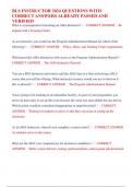
Alveoli
Alveoli are small sacs that is
responsible for gas exchange.
It is surrounded by capillaries.
Alveolar Cells: Pneumocytes
Type 1
- Most common (97% of cells)
- Thin for gas exchange
Type 2
- Produce surfactant
- Can proliferate to other cells - key for regeneration after injury
Club Cells (bronchioles)
- Surfactant
- Responsible for detoxification
Surfactant
When you exhale the alveoli shrinks. The alveoli can collapse and this is called
atelectasis due to decreased efficiency gas exchange.
Surfactant allows alveoli to avoid collapse.
, Surfactants are secreted by type 2 pneumocytes. Mix of lecithins. Especially
dipalmitoylphosphatidylcholine
Fetal Lung Maturity
Lungs are “mature” when enough surfactant are present. Occurs around 35 weeks.
Lecithin-sphingomyelin ratio (L/S ratio)
Both produced equally until approx 35 weeks.
Ratio >2.0 in amniotic fluid suggests lungs mature
Preterm delivery: betamethasone is used to stimulate surfactant production in lungs.
Neonatal Respiratory Distress Syndrome
Hyaline membrane disease. Can lead to atelectasis. There is severe hypoxemia/↑pCO2
(poor ventilation). Poorly responsive to O2 because lungs are collapsed (alveoli) and
intrapulmonary shunting.
Risk Factors
- Prematurity
- Maternal diabetes: high insulin levels decrease surfactant production
- Cesarean delivery: lack of vaginal compression stress leads to reduced fetal
cortisol and reduction in surfactant
There are many complications in NRDS:
1. Bronchopulmonary dysplasia
2. Patent ductus arteriosus (hypoxia keeps shunt open)
3. Retinopathy of prematurity
- Oxygen → free radical formation
- Neovascularization in the retina
- Retinal detachment → blindness
Lobes of the Lung