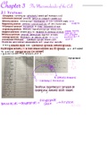
EXAM 2 NOTES PREP
BIO_251 9/17/19
Communication Across a Synapse
1) Action Potential
2) Voltage-gated Ca channels open
3) Calcium triggers exocytosis
4) Neurotransmitter diffuses and binds to receptor
5) Response in cell
- Response terminated by removing neurotransmitter from synaptic cleft
6) Degradation (via enzyme)
7) Reuptake into axon terminal
8) Diffusion away from synapse
Synaptic Delay
- 1-5 msec between arrival of action potential and change in post-synaptic Vm
1) Caused by changes in [Ca2+] and release of neurotransmitter
- NOT related to diffusion of neurotransmitter across synaptic cleft
Postsynaptic Potential
- Change in membrane potential in response to neurotransmitter binding to receptor
1) Produces a graded potential on the membrane of post-synaptic neuron
Excitatory vs. Inhibitory Synapses
1) Excitatory – more likely to have action potential on post-synaptoc cell
a. Depolarization
b. Moves Vm towards threshold
2) Inhibitory – less likely to have action potential
a. Hyperpolarization
b. Moves Vm away from threshold
c. Membrane stabilization
Excitatory Synapses
1) Depolarize postsynaptic cell
a. Brings membrane potential closer to threshold
, i. Depolarization = EPSP = excitatory post-synaptic potential
ii. A type of graded potential
- Can give rise to action potentials on post-synaptic neuron if sufficient to cause membrane
to reach threshold in axon hillock
Inhibitory Synapses
1) Neurotransmitter binds to receptor
2) Channels for either K or Cl open
3) If K channels open:
a. K moves out -> IPSP (inhibitory post-synaptic potential; make ICF more
negative; hyperpolarize the membrane potential)
4) If Cl channels open:
a. Cl moves into cell ->IPSP (makes ICF more negative)
- Can prevent action potentials on post-synaptic neurons; moves membrane potential away
from threshold.
1) A single neuron can receive both excitatory and inhibitory synapses
- So, what determines whether or not a post-synaptic neuron will generate an action
potential?
Neural Integration
- The summing of input from various synapses at the axon hillock of the postsynaptic
neuron will determine whether the neuron will generate action potentials
Convergence of Input as a Factor in Summation
Ex. Two excitatory vs. One inhibitory synapse
Integration can be either spatial (close on membrane) or
temporal (=timing)
- Some neurons receive synapses from 10,000 to
100,000 different neurons – very complex
Neurotransmitters
1) Acetylecholine
2) Biogenic Amines
a. Catecholamines
b. Serotonin
3) Amino Acids
4) Others
Acetylcholine
1) Found in both central and peripheral nervous systems
2) Most abundant neurotransmitter in peripheral nervous system
, - Acetyl CoA + choline -> acetylcholine + CoA
- Synthesized in cytosol of axon terminal
Breakdown of Acetylcholine
Acetylcholine -> acetate + choline
1) Degradation occurs in synaptic cleft
2) Enzyme of degradation = acetylcholinesterase (AChE)
3) Choline recycled and used again
Biogenic Amines
- Derived from amino acids
1) Catecholamines – derived from tyrosine
a. Dopamine
b. Norepinephrine
c. Epinephrine (adrenaline)
2) Serotonin – derived from tryptophan
a. CNS neurotransmitter
b. Main location – brainstem
c. Functions
i. Regulating sleep
ii. Emotions
Mood Enhancing Drugs
1) Many inhibit serotonin re-uptake into axon terminal of neuron pathways involved with
mood.
a. Ex. Prozac, Paxil, Zoloft
2) Increased levels of serotonin remain in synaptic cleft and continue to stimulate post-
synaptic neurons
3) Used to treat depression (however, in some people cause a paradoxical worsening of
depression)
Amino Acid Neurotransmitters
- In central nervous system
1) Aspartate (Excitatory – produce EPSP’s)
2) Glutamate (same^)
3) Glycine (same as below)
4) GABA (Inhibitory – produce IPSP’s)
Other Neurotransmitters
1) NO = Nitric Oxide
a. Gas
b. Produced by nitric oxide synthetase (enzyme)
Two Interconnected Parts to Nervous System
1) Central Nervous System
a. Brain and spinal cord (integrating and command center)
2) Peripheral Nervous System
, a. Outside the CNS
b. Consists of nerves extending from brain and spinal cord
c. Peripheral nerves link all regions of the body to the CNS
d. Includes Somatic, Autonomic, and Sensory
Part 1: Autonomic Nervous System
- Also known as the “visceral” nervous system because innervates most internal organs
(heart, digestive system, lungs, etc)
- Controls smooth muscle, cardiac muscle, glands, and adipose (fat cells)
- TWO divisions: Sympathetic and Parasympathetic Nervous System
Dual Innervation of the Autonomic Nervous System
- Both branches of the autonomic nervous system innervate most organs
- Primary function – regulate organs to maintain homeostasis
- Parasympathetic and sympathetic activities tend to oppose each other
Anatomy of the Autonomic Nervous System
1) Two neurons from CNS to organs (connected in series)
a. Preganglionic neuron
b. Postganglionic neuron
2) Autonomic Ganglia
a. Synapse between pre- and postganglionic neurons occurs in ganglia (collection of
neuron cell bodies outside CNS)
3) Axons of postganglionic neurons innervate target organs
Anatomy of Autonomic Pathwaves
Sympathetic Nervous System
- Preganglionic neurons originate in thoracic and lumbar regions of spinal cord
- General Anatomy
o Short preganglionic neurons to sympathetic chain ganglia (along vertebral
column)
o Most ganglia linked together in sympathetic chain
o Long postganglionic neurons from chain to effector organs