Procedure:
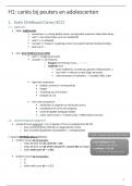
Aim:
Matched pair design
To investigate whether there was d
Sample
brain functioning in a group of mu
Two groups- criminals and control group control participants.
41 participants (39 males and 2 female)
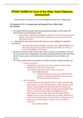
Prefrontal cortex
All were convicted for murder or manslaughter
Amygdala
Claimed “not guilty by reason of insanity” NGRIs Biological Hippocampus
6 schizophrenia Classic study Thalamus
Corpus callosum
23 organic brain damage or head injury
3 substance abusers Raine et al
2 affective disorders (1997) Results:
Control group matched on age and gender ( mean age Brain dysfunction in NGRI group
of murderers 34.4) implicated in violent behaviours.
Conclusion: Murderers showed:
Method Brain differences have been • Lower activity in prefrontal c
All participants in this group remained medication-free associated with many behavioural • Lower activity in parietal cort
for two weeks prior to PET scan changes. • Higher activity in the occipita
Suggesting abnormal activity could • Identical activity in temporal
Given continuous performance task
result in criminals being unable to • Lower activity in corpus callo
Blurred numbers to focus on modify behaviour • Asymmetrical behaviour in a
The participants were injected with glucose. Certain differences make violence
more likely to occur.
32 minutes after a PET scan was conducted to observe
metabolic rate in different areas of the brain. (activity
levels)
Aim:
Matched pair design
To investigate whether there was d
Sample
brain functioning in a group of mu
Two groups- criminals and control group control participants.
41 participants (39 males and 2 female)
Prefrontal cortex
All were convicted for murder or manslaughter
Amygdala
Claimed “not guilty by reason of insanity” NGRIs Biological Hippocampus
6 schizophrenia Classic study Thalamus
Corpus callosum
23 organic brain damage or head injury
3 substance abusers Raine et al
2 affective disorders (1997) Results:
Control group matched on age and gender ( mean age Brain dysfunction in NGRI group
of murderers 34.4) implicated in violent behaviours.
Conclusion: Murderers showed:
Method Brain differences have been • Lower activity in prefrontal c
All participants in this group remained medication-free associated with many behavioural • Lower activity in parietal cort
for two weeks prior to PET scan changes. • Higher activity in the occipita
Suggesting abnormal activity could • Identical activity in temporal
Given continuous performance task
result in criminals being unable to • Lower activity in corpus callo
Blurred numbers to focus on modify behaviour • Asymmetrical behaviour in a
The participants were injected with glucose. Certain differences make violence
more likely to occur.
32 minutes after a PET scan was conducted to observe
metabolic rate in different areas of the brain. (activity
levels)