Neuroanatomy I & II
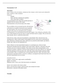
The neuron
Senses changes in the environment, communicate these changes to other neurons and command the
body's responses to these sensations.
Consist of:
- Dendrite
- Soma (cell body containing the organelles)
- Axon, which ends in synapses
An action potential travels along axons and their presence is
signaled to the next neuron at the synapse.
Neurotransmitters convey the message at the synapse.
The axon hillock is the area formed from the cell body/soma
of the neuron as it extends to become the axon. It precedes
the initial segment. The received action potentials that are
summed in the neuron are transmitted to the axon hillock
for the generation of an action potential from the initial segment. Axon collaterals are branches of the
axon. At the terminal site, the axon comes in contact with other neurons (synapse). This can also have
many branches, then it is called the terminal arbor.
In the nervous system, axons may be myelinated, or unmyelinated. This is the provision of an
insulating layer, called a myelin sheath. In the peripheral nervous system axons are myelinated by
glial cells known as Schwann cells. In the central nervous system the myelin sheath is provided by
another type of glial cell, the oligodendrocyte.
Nodes of Ranvier are short unmyelinated segments of a myelinated axon, which are found
periodically interspersed between segments of the myelin sheath. Therefore, at the point of the node
of Ranvier, the axon is reduced in diameter. These nodes are areas where action potentials can be
generated. In saltatory conduction, electrical currents produced at each node of Ranvier are conducted
with little attenuation to the next node in line, where they remain strong enough to generate another
action potential. Thus in a myelinated axon, action potentials effectively "jump" from node to node,
bypassing the myelinated stretches in between, resulting in a propagation speed much faster than even
the fastest unmyelinated axon can sustain.
Classifying neurons
Unipolar: a neuron with a single neurite (axon/dendrite)
Bipolar: two neurites
Multipolar: three or more, most of the brain neurons are multipolar
Sensory: have neurites in the sensory surface
Motor: axons that form synapses with the muscles and command movement
Interneurons: connection with another neuron
Neuroglia
,Glia contributes to brain function mainly by isulating, supporting and nourishing neighboring
neurons.
Neuroglia - Central nervous system
Astrocytes
Astrocytic extensions cover the entire outside surface of the brain
Glia limitans perivascular
- Surrounds all blood vessels
- Part of the blood-brain barrier (controls which enters the brain)
Glia limitans superficialis
- Borders on subarachnoid space
- Part of the brain-liquor barrier
Astrocytic extensions cover all uncovered surfaces of the neuronal somata, dendrites and
(unmyelinated axons).
- Structural support
- Control of ECF by uptake of K+ and neurotransmitters
Oligodendrocytes
Form the myelin sheaths in CNS. Unmyelinated axons are covered by astrocytes. These are needed to
increase the speed of the action potential.
, Ependymal cells
Squamous to the columnar epithelial lining of the ventricles.
- May be ciliated; cilia beat to circulate CSF
- Form a permeable barrier between CSF and ECF, so the astrocytes
will be present underneath
- Form the choroid plexus
- Production of cerebrospinal fluid (CSF)
- Absorption of waste and unnecessary solutes from SCF
Microglial cells
Mononuclear phagocytes. They enter the brain and clean up dead neurons (waste disposal). These
cells are very important and can cause severe diseases. The progenitor cells are present everywhere.
Neuroglia - Peripheral nervous system
Satellite cells
Similar in function to the astrocytes. They cover the cell body of the neuron to protect.
Swann cells
Cover all axons in PNS.
Schwann cells cover all axons in the PNS. There are myelinating Schwann
cells which form myelin sheats bu also non-myelinating Schwann cells,
which form an envelope for non-myelinated axons (remak bundles).