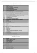
Use of this content is subject to the Terms and Conditions of the Evolve web site.
A2
Biology
STRUCTURE AND FUNCTION OF NORMAL CELLS
STRUCTURE AND FUNCTION OF NORMAL CELLS
All normal cells of the human body have some common features and consist of the same basic components.
These include the nucleus, the cytoplasm, and the cell (plasma) membrane (Figure 1-1).
Figure 1-1 Normal cells have a nucleus and a cytoplasm. On the outside, the cell is delimited by a plasma
membrane. In the cytoplasm, there are organelles, such as mitochondria, smooth and rough endoplasmic
reticulum (SER and RER, respectively), Golgi apparatus, and lysosomes.
Nucleus
The nucleus is the essential part of most living cells. It consists of nucleic acids, such as deoxyribonucleic
acid (DNA) and ribonucleic acid (RNA), and nuclear proteins. In resting cells, these components are arranged
into aggregates known as chromatin and a specialized organelle composed primarily of RNA known as the
nucleolus. In the dividing of cells—that is, during mitosis—the chromatin is restructured and the strands of
DNA condense into chromosomes. The resting cells have a nuclear membrane, which delimits the nucleus
from the cytoplasm. This membrane disappears in mitosis and reappears after cell division is completed.
The DNA of the nucleus contains essential genetic material that is identical for all cells of an individual body.
This genetic material consists of genes that are differentially expressed in various tissues and organs.
Differential expression of genes allows the cells to assume unique features in various tissues and organs and
to perform specialized functions. Such cells are called differentiated, in contrast to embryonic cells, which
have not undergone specialization and which are therefore termed undifferentiated.
The genetic information encoded in the DNA is transcribed into the nuclear RNA. From the nuclear RNA, the
message is transmitted by transfer RNA (tRNA) and messenger RNA (mRNA) into the cytoplasm (Figure 1-
2). The ribosomal RNA (rRNA) serves as a template for translating the genetic messages into proteins.
Protein synthesis is essential for the maintenance of life. Proteins are needed for cellular growth, replication,
,metabolism, respiration, and other essential functions. Proteins also act as structural elements, maintaining the
cell's shape and the internal organization of the cytoplasm. None of these elementary functions (and many
others that we mention later) would be possible without the nucleus, which acts as the main overseer of all
critical cytoplasmic events. All human cells, except the red blood cells and platelets, need a nucleus for
survival.
Figure 1-2 Transcription and translation by RNA of the genetic code stored in the DNA leads to protein
synthesis on ribosomes. mRNA, messenger RNA; RER, rough endoplasmic reticulum; tRNA, transfer RNA.
Cytoplasm
All cells have cytoplasm. However, the amount of cytoplasm and its structure vary from one cell to another. In
embryonic cells, the cytoplasm is scant and contains few organelles. In specialized, highly differentiated cells,
such as liver or kidney cells, the cytoplasm is more abundant and is replete with organelles. The ratio of the
nucleus to the cytoplasm, the so-called nucleocytoplasmic (N : C) ratio, is high in undifferentiated embryonic
cells and much lower in differentiated cells of adult tissues. As we shall see later, many tumor cells are also
undifferentiated and have a high N : C ratio.
The principal cytoplasmic organelles are the mitochondria, ribosomes, endoplasmic reticulum, Golgi
apparatus, and lysosomes. In addition to these, some cells have organelles for specialized functions. For
example, muscle cells have myofilaments composed of actin and myosin, which are essential for contraction;
glandular cells have secretory granules, which contain enzymes or mucus destined for excretion. Furthermore,
it is important to note that the cytoplasmic ground substance of all cells consists of an amorphous matrix
called hyaloplasm and a fibrillar meshwork called cytoskeleton. Each cell is also enclosed by an outer plasma
membrane, which forms the border between one cell and other cells or the extracellular spaces. This
membrane, which is semipermeable, must remain intact to preserve the viability of the cell.
Mitochondria.
Mitochondria are double-membrane–bound cytoplasmic organelles, involved primarily in the generation of
energy (see Figure 1-1). Hence, mitochondria are rich in oxidative enzymes. These enzymes (e.g., cytochrome
oxidase) are attached to the double membrane that encloses each mitochondrion and to the cristae that are
seen by electron microscopy on the inside of cross-sectioned mitochondria. Energy generated by the
mitochondria is essential for all other cellular functions. Cells with complex functions, such as liver cells and
nerve cells, require a considerable amount of energy and therefore contain numerous mitochondria. By
comparison, undifferentiated cells, including many malignant tumor cells, have few mitochondria.
Ribosomes.
Ribosomes are small granules composed of RNA. They may be arranged into aggregates that float freely in
the cytoplasm, called polysomes or free ribosomes, or they may be attached to the membranes of the rough
endoplasmic reticulum (RER). The ribosomes are involved in protein synthesis. Structural proteins and
enzymes needed for the maintenance of basic cell functions (“proteins for internal purposes”) are synthesized
on the free ribosomes. Those intended for excretion (“export or luxury proteins”) are synthesized on the RER
and discharged from the cells through the cisternae lined by the membranes of the RER.
Endoplasmic Reticulum.
The endoplasmic reticulum is a meshwork of membranes that are in continuity with the outer plasma
membranes on one side and the nuclear membrane on the other (Figure 1-3). With use of electron microscopy,
, one can distinguish two forms of endoplasmic reticulum: the RER and the smooth endoplasmic reticulum
(SER). As stated earlier, the RER is the site of protein synthesis. SER has complex functions, the most
important of which are the catabolism (i.e., metabolic degradation) of drugs, hormones, and various nutrients
and the synthesis of steroid hormones. SER is therefore most prominent in liver cells, known for their
complex catabolic functions. The hormone-secreting gonadal cells of the testis and ovary and the
adrenocortical cells that synthesize steroid hormones (e.g., estrogens, androgens, and corticosteroids) also
have prominent SER.
Figure 1-3 Endoplasmic reticulum. It consists of rough endoplasmic reticulum (RER) arranged into
ribosome-studded cisternae and vesicles of smooth endoplasmic reticulum (SER).
Golgi Apparatus.
The Golgi apparatus is a synthetic organelle adjacent to the nucleus (see Figure 1-3). Its tubules and flattened
cisternae, which are its main components, give rise laterally to vesicles. The vesicles arising from the concave
side of the Golgi apparatus—the maturing surface—become secretory granules, lysosomes, and specialized
structures, such as melanosomes. Melanosomes are the melanin-containing organelles of pigmented cells
(melanocytes) in the skin and eye. The convex face is in continuity with the endoplasmic reticulum. Many
proteins synthesized in the endoplasmic reticulum pass through the Golgi apparatus, where they are
biochemically modified before being packaged into secretory granules or lysosomes. Glycoproteins and
lipoproteins (i.e., proteins linked to a carbohydrate or lipid) are formed in the Golgi apparatus. These complex
proteins are then incorporated into the internal cell membranes (e.g., endoplasmic reticulum) or the outer
plasma membrane or are secreted from the cell.
Lysosomes.
Lysosomes are membrane-bound digestive cytoplasmic organelles that are rich in lytic enzymes. The
lysosomes originate as small vesicles budding from enzymes on the maturing face of the Golgi apparatus
(Figure 1-4). These primary lysosomes contain acid hydrolases, which are digestive enzymes that are
maximally active in an acidic milieu (i.e., at low pH levels). Under normal circumstances, the lytic enzymes
are tightly enclosed by a lysosomal outer membrane and do not harm the cell. Even if some lysosomal content
is spilled into the cytoplasm, the acid hydrolases would cause little damage in normal cytoplasm, which has a
neutral pH. However, if the cell is injured and the pH of the cytoplasm becomes acidic, enzymes released
from the lysosomes could cause damage.
Figure 1-4 Lysosomes. Primary (1°) lysosomes, which originate from the Golgi apparatus, give rise to
heterophagosomes and autophagosomes. Undigested material in phagosomes is extruded from the cell or
remains in the cytoplasm as lipofuscin-rich residual bodies. RER, rough endoplasmic reticulum.
The primary lysosomes fuse with other cytoplasmic vesicles to form secondary lysosomes. Typically, they
fuse with the absorptive vesicles originating from the invaginated plasma membrane to form secondary
lysosomes, which are also called heterophagosomes. Secondary lysosomes that are involved in the
autodigestion of a cell's own organelles are called autophagosomes. The digestive enzymes in secondary
lysosomes degrade the material enclosed within its membrane. The metabolites obtained through this
intracellular digestion are reutilized within the cell's cytoplasm. The undigested residues are extruded from the
cytoplasm into the extracellular spaces by reverse endocytosis or exocytosis, a process that is colloquially