Topic 8: Grey Matter
Brain Development, Drugs and Disease
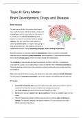
Brain structure
The brain can be divided into specific regions which
have specific functions. Most of our brain is made up of
the cerebrum, which is found at the top of the brain. It
is divided into two cerebral hemispheres joined
together by a band of nerve fibres called the corpus
callosum. The thin, outer layer of the cerebrum is
called the cerebral cortex. It is highly folded, giving it a
really large surface area. The cerebrum is involved in
‘higher-brain functions’, such as processing language, vision, thinking and emotions.
Below the cerebrum is a structure called the hypothalamus, which is involved in homeostatic
responses such as maintaining body temperature (thermoregulation). It also produces hormones
that control the pituitary gland, which is found just beneath the hypothalamus.
The cerebellum is a leaf-shaped structure found towards the back of the brain. It is positioned
underneath the cerebrum and is highly folded. It plays an important role in movement and balance.
Things like learning to ride a bike or the movement involved in writing will involve a large input from
the cerebellum.
Right at the base of the brain and above the spinal cord is a structure called the medulla oblongata.
This is involved in unconscious processes, such as the regulation of breathing rate and heart rate.
CT scans
• Computed tomography (CT) scans are images of the brain which can be taken to view the
structure of the brain and to assess whether brain damage has occurred.
• Use X-ray radiation to produce a cross-section image of the brain.
• Denser brain structures absorb more X-ray radiation and show up as darker regions on the
image.
• Useful for diagnosing medical conditions because they can indicate areas of disease or
damage. For example, bleeding in the brain is visible on CT scans because blood had a
different density to brain tissue.
, Although you cannot use CT scanners to work out the function of different brain regions directly, you
can infer the functions of different brain regions by matching a patient’s symptoms with areas of
brain damage. For example, if a CT scan of a person with dementia shows damage to the cerebrum,
this indicates that the cerebrum plays a role in the consolidation of memories.
MRI scans
• Magnetic resonance imaging (MRI) scanners use radio waves in the presence of a strong
magnetic field to produce cross-sectional images of the brain.
• Higher quality and have a higher resolution than CT scans.
• Also used in medical diagnosis because they can be used to visualise damaged or diseased
parts of the brain. For example, MRI scans can be used to indicate the presence of a brain
tumour, which shows up as a lighter colour on an MRI scan.
• Doctors can determine the size and location of the tumour and decide on the best treatment
option to take. Just like CT scanning, the function of brain regions can be inferred by
matching up a patient’s symptoms with areas of brain damage.
fMRI scans
• Function magnetic resonance imaging (fMRI) scanners show which areas of the brain are
active by detecting changes in blood flow.
• Brain regions which are stimulated will be respiring more, so more blood will be flowing to
these regions (to deliver more glucose and oxygen).
• Oxygenated blood produces a stronger signal in a magnetic field compared to
deoxygenated blood, so these areas show up on the fMRI scan.
fMRI scans are similar to MRI scans but they can also be used to research the function of different
brain structures. For example, a person inside the scanner may be asked to look at images of
different faces. The areas of the brain which light up on the fMRI scan will indicate the brain regions
which are involved in facial recognition. fMRI scans are also used in medical diagnosis since they
show damaged and diseased parts of the brain.
PET scans
• Like fMRI, positron emission tomography (PET) scans also show which regions of the
brain are activated at any given time, but they use radioactive tracers such as radioactively
labelled glucose.
• The radioactively labelled glucose will accumulate in parts of the brain which are respiring
more and will produce a stronger signal on the PET scan.
• Just like MRI scans, PET scans have a high resolution and high quality.
Brain Development, Drugs and Disease
Brain structure
The brain can be divided into specific regions which
have specific functions. Most of our brain is made up of
the cerebrum, which is found at the top of the brain. It
is divided into two cerebral hemispheres joined
together by a band of nerve fibres called the corpus
callosum. The thin, outer layer of the cerebrum is
called the cerebral cortex. It is highly folded, giving it a
really large surface area. The cerebrum is involved in
‘higher-brain functions’, such as processing language, vision, thinking and emotions.
Below the cerebrum is a structure called the hypothalamus, which is involved in homeostatic
responses such as maintaining body temperature (thermoregulation). It also produces hormones
that control the pituitary gland, which is found just beneath the hypothalamus.
The cerebellum is a leaf-shaped structure found towards the back of the brain. It is positioned
underneath the cerebrum and is highly folded. It plays an important role in movement and balance.
Things like learning to ride a bike or the movement involved in writing will involve a large input from
the cerebellum.
Right at the base of the brain and above the spinal cord is a structure called the medulla oblongata.
This is involved in unconscious processes, such as the regulation of breathing rate and heart rate.
CT scans
• Computed tomography (CT) scans are images of the brain which can be taken to view the
structure of the brain and to assess whether brain damage has occurred.
• Use X-ray radiation to produce a cross-section image of the brain.
• Denser brain structures absorb more X-ray radiation and show up as darker regions on the
image.
• Useful for diagnosing medical conditions because they can indicate areas of disease or
damage. For example, bleeding in the brain is visible on CT scans because blood had a
different density to brain tissue.
, Although you cannot use CT scanners to work out the function of different brain regions directly, you
can infer the functions of different brain regions by matching a patient’s symptoms with areas of
brain damage. For example, if a CT scan of a person with dementia shows damage to the cerebrum,
this indicates that the cerebrum plays a role in the consolidation of memories.
MRI scans
• Magnetic resonance imaging (MRI) scanners use radio waves in the presence of a strong
magnetic field to produce cross-sectional images of the brain.
• Higher quality and have a higher resolution than CT scans.
• Also used in medical diagnosis because they can be used to visualise damaged or diseased
parts of the brain. For example, MRI scans can be used to indicate the presence of a brain
tumour, which shows up as a lighter colour on an MRI scan.
• Doctors can determine the size and location of the tumour and decide on the best treatment
option to take. Just like CT scanning, the function of brain regions can be inferred by
matching up a patient’s symptoms with areas of brain damage.
fMRI scans
• Function magnetic resonance imaging (fMRI) scanners show which areas of the brain are
active by detecting changes in blood flow.
• Brain regions which are stimulated will be respiring more, so more blood will be flowing to
these regions (to deliver more glucose and oxygen).
• Oxygenated blood produces a stronger signal in a magnetic field compared to
deoxygenated blood, so these areas show up on the fMRI scan.
fMRI scans are similar to MRI scans but they can also be used to research the function of different
brain structures. For example, a person inside the scanner may be asked to look at images of
different faces. The areas of the brain which light up on the fMRI scan will indicate the brain regions
which are involved in facial recognition. fMRI scans are also used in medical diagnosis since they
show damaged and diseased parts of the brain.
PET scans
• Like fMRI, positron emission tomography (PET) scans also show which regions of the
brain are activated at any given time, but they use radioactive tracers such as radioactively
labelled glucose.
• The radioactively labelled glucose will accumulate in parts of the brain which are respiring
more and will produce a stronger signal on the PET scan.
• Just like MRI scans, PET scans have a high resolution and high quality.