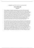
DVT probability
(Wells’ score)
Likely Unlikely
Compression Hypercoagulation Disorders
US D-dimer
- + - +
Compression
D-dimer Treat No DVT US
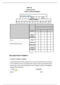
Venous Thromboembolism (VTE)
Stasis
Post-thrombotic syndrome:
• Development of chronic venous stasis signs/symptoms 2/2 DVT
• Thrombus formation à inflammatory response in superficial or deep vein • Pain, venous dilation, edema, pigmentation, venous ulcers
• Superficial thrombophlebitis • Villalta score: clinical severity
• Deep vein thrombosis (DVT) • Tx: elevation, exercise, compression stockings, intermittent pneumatic
• Pulmonary embolism (PE) compression therapy, skin/wound care
Pathophysiology: Endothelial Ddx elevated D-dimer:
- + - + 1. Endothelial damage à induces hemostasis
2. Venous stasis à inhibits clearance and dilution of coag factors
Hypercoagulability injury • Arterial thromboembolic dz (MI, CVA, Afib)
• Venous thromboembolic disease (DVT, PE)
• DIC
3. Hypercoagulability = more likely to thrombose • Preeclampsia and eclampsia
• Increases with age, surgery, trauma, neoplasm, blood dyscrasias, prolonged immobilization, hormones (E), APS, heart failure • Abnormal fibrinolysis, use of thrombolytic agents
• CVD, CHF
DVT • Severe infection/sepsis/inflamm
Clinical presentation: • Surgery/trauma
Serial US • Unilateral leg edema, erythema, warmth, tenderness, palpable cord (thrombosed vein) • SIRS
No DVT (5-7 d) No DVT Treat • Phlegmasia alba dolens (white appearance), phlegmasia cerula dolens (acute pain and edema) with massive thrombosis
• Sickle cell vasoocclusive episode
• Severe liver dz
• Homan’s sign (pain with dorsiflexion) (unreliable) • Malignancy
Ddx: muscles strain/tear, lymphangitis/lymph obstruction, venous valve insufficiency, ruptured popliteal cyst, cellulitis, arterial occlusive dz • Renal disease
• Normal pregnancy
• Venous malformation
INVESTIGATIONS
• D-dimer Test Principle Readout Pros/Cons
• Doppler US* D-dimer breakdown product of fibrin positive/negative sensitive but not specific; cannot be used alone
- + • MRI
Compression detects “non-compression” of a positive/negative sensitive for proximal; one negative does not rule out
• Impendence plethysmography ultrasound venous segment DVT
• Venography
• CTPA, V/Q scan if PE suspected V/Q scan looks for ventilation/perfusion normal/low/intermediate/high probability preferred in younger pts with normal CXR
mismatch in lungs *normal excludes PE more “non-diagnostic” results
CTPA rapid CT with thin slices, bolus of negative/positive/technically inadequate preferred in individuals with a positive D-dimer
dye and radiation *negative can rule out a PE more results of unknown clinical significance
No DVT Treat contraindicated if renal issues
may catch more clots that aren’t clinically relevant
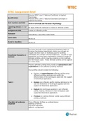
, Coagulation Cascade
(Wells’ score)
Likely Unlikely
Compression Hypercoagulation Disorders
US D-dimer
- + - +
Compression
D-dimer Treat No DVT US
Venous Thromboembolism (VTE)
Stasis
Post-thrombotic syndrome:
• Development of chronic venous stasis signs/symptoms 2/2 DVT
• Thrombus formation à inflammatory response in superficial or deep vein • Pain, venous dilation, edema, pigmentation, venous ulcers
• Superficial thrombophlebitis • Villalta score: clinical severity
• Deep vein thrombosis (DVT) • Tx: elevation, exercise, compression stockings, intermittent pneumatic
• Pulmonary embolism (PE) compression therapy, skin/wound care
Pathophysiology: Endothelial Ddx elevated D-dimer:
- + - + 1. Endothelial damage à induces hemostasis
2. Venous stasis à inhibits clearance and dilution of coag factors
Hypercoagulability injury • Arterial thromboembolic dz (MI, CVA, Afib)
• Venous thromboembolic disease (DVT, PE)
• DIC
3. Hypercoagulability = more likely to thrombose • Preeclampsia and eclampsia
• Increases with age, surgery, trauma, neoplasm, blood dyscrasias, prolonged immobilization, hormones (E), APS, heart failure • Abnormal fibrinolysis, use of thrombolytic agents
• CVD, CHF
DVT • Severe infection/sepsis/inflamm
Clinical presentation: • Surgery/trauma
Serial US • Unilateral leg edema, erythema, warmth, tenderness, palpable cord (thrombosed vein) • SIRS
No DVT (5-7 d) No DVT Treat • Phlegmasia alba dolens (white appearance), phlegmasia cerula dolens (acute pain and edema) with massive thrombosis
• Sickle cell vasoocclusive episode
• Severe liver dz
• Homan’s sign (pain with dorsiflexion) (unreliable) • Malignancy
Ddx: muscles strain/tear, lymphangitis/lymph obstruction, venous valve insufficiency, ruptured popliteal cyst, cellulitis, arterial occlusive dz • Renal disease
• Normal pregnancy
• Venous malformation
INVESTIGATIONS
• D-dimer Test Principle Readout Pros/Cons
• Doppler US* D-dimer breakdown product of fibrin positive/negative sensitive but not specific; cannot be used alone
- + • MRI
Compression detects “non-compression” of a positive/negative sensitive for proximal; one negative does not rule out
• Impendence plethysmography ultrasound venous segment DVT
• Venography
• CTPA, V/Q scan if PE suspected V/Q scan looks for ventilation/perfusion normal/low/intermediate/high probability preferred in younger pts with normal CXR
mismatch in lungs *normal excludes PE more “non-diagnostic” results
CTPA rapid CT with thin slices, bolus of negative/positive/technically inadequate preferred in individuals with a positive D-dimer
dye and radiation *negative can rule out a PE more results of unknown clinical significance
No DVT Treat contraindicated if renal issues
may catch more clots that aren’t clinically relevant
, Coagulation Cascade