Notes: Please adhere to the guidelines explained in Histology 101,
which can be found under the Important Lab Documents drop-
down menu on the Lab Resources tab of our OWL site. Marks may
be deducted if you do not read and follow these instructions.
Lab Assignment 1: Microscopy & Histology Techniques
1. Of the following structures, which can be positively identified with a light microscope? Transmission
electron microscope? Briefly explain your reasoning below the table. (2.5 marks)
Transmission Electron
Light Microscope
Microscope
( anbe dentifred becunhe idenlity bloodcel
Red blood cell (7µm) 7 , um ) Tem Lonld becuase 3 1
" D 21 µ m 7 mm .
0
nm
where o 21 µm is
Liyhy microstope ' s
resolulom where . m is TEM resolntion
be identified belause
Plasma membrane (10nm) cunl
Liyh * miroscope ) 7 em Loubd identit , plasma membrun 은 IDnm 3 1 Dnm
resolulion is D 21 µm , 1 1 tor lonm
.
are where 1 onm is TEM resolution .
definubble befunn
Resolution
* : minimul
tw
. poil 정
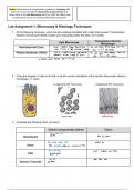
2. Using the diagram of cilia on the left, circle the correct orientation of the section above each electron
micrograph. (1 mark)
Longitudinal OR Transverse Longitudinal OR Transverse
frunsrerle seation loyiladimalsection
https://med.libretexts.org/Bookshelves/Anatomy_and_Physiology/Human_Anatomy_(OERI)/02%3A_Cellular_Level_of_Organization/2.02%3A_The_Cell_Membrane
3. Complete the following chart. (4 marks)
Cellular component(s) stained Colour
staini mudeic usid
RWA
IUK
blue dye Esfain )
Hematoxylin
,
,
stains positively Ehurghd parficler Red /pink dye
Eosin Proteiny ,
Eond 5 luih Eytopluym and intrumembramius profeiss
Stain , Earbohydrules muzantu Lolor < slaind
bright
PAS glyolipid ' yluose
siains Iipids Danse blask wlor
Osmium Tetroxide lipd biluyers
which can be found under the Important Lab Documents drop-
down menu on the Lab Resources tab of our OWL site. Marks may
be deducted if you do not read and follow these instructions.
Lab Assignment 1: Microscopy & Histology Techniques
1. Of the following structures, which can be positively identified with a light microscope? Transmission
electron microscope? Briefly explain your reasoning below the table. (2.5 marks)
Transmission Electron
Light Microscope
Microscope
( anbe dentifred becunhe idenlity bloodcel
Red blood cell (7µm) 7 , um ) Tem Lonld becuase 3 1
" D 21 µ m 7 mm .
0
nm
where o 21 µm is
Liyhy microstope ' s
resolulom where . m is TEM resolntion
be identified belause
Plasma membrane (10nm) cunl
Liyh * miroscope ) 7 em Loubd identit , plasma membrun 은 IDnm 3 1 Dnm
resolulion is D 21 µm , 1 1 tor lonm
.
are where 1 onm is TEM resolution .
definubble befunn
Resolution
* : minimul
tw
. poil 정
2. Using the diagram of cilia on the left, circle the correct orientation of the section above each electron
micrograph. (1 mark)
Longitudinal OR Transverse Longitudinal OR Transverse
frunsrerle seation loyiladimalsection
https://med.libretexts.org/Bookshelves/Anatomy_and_Physiology/Human_Anatomy_(OERI)/02%3A_Cellular_Level_of_Organization/2.02%3A_The_Cell_Membrane
3. Complete the following chart. (4 marks)
Cellular component(s) stained Colour
staini mudeic usid
RWA
IUK
blue dye Esfain )
Hematoxylin
,
,
stains positively Ehurghd parficler Red /pink dye
Eosin Proteiny ,
Eond 5 luih Eytopluym and intrumembramius profeiss
Stain , Earbohydrules muzantu Lolor < slaind
bright
PAS glyolipid ' yluose
siains Iipids Danse blask wlor
Osmium Tetroxide lipd biluyers