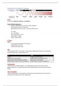
Introduction to Medical Imaging
X-RAY
X-rays can be deflected, scattered, and absorbed
Factors Affecting Appearance
1. Density – determines how much X-ray will be absorbed
Radio-opaque: white on X-ray (bone)
Radio-lucent: black on X-ray (air – absorb very little X-rays)
Air – Black
Fat – Grey/Black
Soft Tissues/Organs – Grey
Calcium/Bone – White
Metal – Intense White
CT SCAN
Planes
- Transverse Plane/Axial Plane (from above)
- Sagittal Plane (from side)
- Coronal Plane (from front)
MRI
Contraindication of MRI – pacemakers, insulin pumps, implanted hearing aids, neurostimulators,
intracranial metal clips, metallic bodies in eye
CT MRI
X-rays Magnet and radio waves
Better used in trauma cases Better used to examine spinal canal
Broken bones and vertebrae more visible Brain tumour more visible
Better image of organs in chest and abdomen Better image of tendons and ligaments
ULTRASOUND
*Does not involve ionizing radiation – uses high frequency sound waves, and is real-time
Doppler Ultrasound – measure blood flow in vessels (depending on sound frequency in flowing
blood)
USE OF CONTRAST
*Barium (swallowed for GIT examinations), iodinated contrast (administered intravascularly or
intrathecally to outline arteries, veins, spinal cord)
,Normal Radiograph – Spine
When describing X-RAY, must mention LEFT/RIGHT + TYPE OF VIEW (AP/PA/LATERAL) + BODY
PART (SHOULDER JOINT/ANKLE JOINT)
*Parts of vertebrae – cervical (7), thoracic (12), lumbar (5), sacrum (5), coccyx (4)
*Parts of vertebral body – transverse process, spinous process
*Where spinal cord ends – L1 or L2
*In spine trauma, CT is highly recommended if there is suspicion of injury / MRI is recommended for
spinal cord and nerve injury
*Spondylosis, osteomyelitis, discitis, abscess, spinal cord tumour – use MRI
CERVICAL
Function – support the weight of head
*C1 – atlas (ring-shaped, for nodding), C2 – axis (peg-shaped, odontoid process, for shaking head)
Projections
1. Antero-Postero View – vertebral bodies and intervertebral spaces
2. Lateral – zygapophyseal joints, soft tissue structure around, spinous processes, AP
relationship of vertebral bodies
3. Odontoid – C1 C2 peg projection
4. AP Oblique – show intervertebral foramina further from image receptor
5. PA Oblique – show intervertebral foramina closer to image receptor
6. Cervicothoracic View – modified lateral projection of C7 and T1 junction
7. Flexion-Extension Lateral – to assess spinal stability
THORACIC
Function – hold rib cage and protect heart & lungs
Projections
1. AP View – performed erect / can see intervertebral joints
2. Lateral View – can see posterior spinous processes / intervertebral joints / neural foramen
(ideal for suspected fractures or dislocations)
, *Identify rib number (anterior and posterior), vertebra number, clavicle, pedicle, intervertebral
foramen & disc
LUMBAR
Function – bear weight of body
Projections
1. PA/AP View – view entire spine (including T11-12 and sacrum, performed erect)
2. Lateral View – pedicles, lumbar vertebral bodies, facet joints (for fractures)
3. Horizontal Beam Lateral – for trauma
4. Oblique View – view articular facets and pars interarticularis
5. Flexion-Extension – to assess spinal stability
*Identify rib number, vertebra number, spinous & transverse process, psoas muscle, sacroiliac joint,
ala of sacrum, intervertebral disc
*Indications – scoliosis, spine metastases (‘winking owl’ at pedicle)
SACRUM
Function – connect spine to iliac bones
*Identify pubic symphysis, sacroiliac joint, ala of sacrum
COCCYX
Function – provide attachment of ligaments and muscles of pelvic floor
INTERVERTEBRAL DISCS
Function – keep bones from rubbing together
1. Nucleus – gel-filled center
2. Annulus – tough fibrous outer ring
FACET JOINTS
Function – allow back motion
*Each vertebra has 4 facet joints, superior facets (connect above) & inferior facets (connect below)
SPINAL CORD
*Measures 42-45cm x 1cm in diameter
*Extends as continuation of medulla oblongata from corticomedullary junction at the foramen
magnum down to tip of conus medullaris at L2 -> at L2 it will change into conus medullaris -> cauda
equina (L2 to Coccyx 1)
*31 nerve roots in total – 8C, 12T, 5L, 5S, 1C
COVERINGS AND SPACES
*Spinal cord is covered by 3 membranes – pia / arachnoid / dura mater (outermost)
*Space between pia and arachnoid mater – wide subarachnoid space (contains CSF)