Cardiac-Dysrhythmia - Cheat sheet for dysrhythmias.
, Rhythm Clinical Associations ECG Characteristics Clinical Significance Treatment S
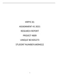
Sinus Bradycardia −Normal in some aerobic athletes -HR: <60 bpm −Depends on how pt hemodynamically −Atropine (anticholinergic) if
−Conduction path same as NSR and some pts during sleep −Rhythm: regular tolerates symptomatic
−SA node fires at <60 bpm −Carotid sinus massage, Vasalva −P wave: normal, before each −S/sx of symptomatic Bradycardia: −Possible pace maker
−Symptomatic− HR <60 resulting maneuver, Hypothermia, QRS pale, cool skin; hypotension; −D/t drugs: d/c, reduce dose, hold
in symptoms (chest pain, syncope Increased intraocular pressure, −PR Int: normal weakness; angina; dizziness or
Vegal stimulation −QRS: normal shape/duration syncope; confusion or disorientation;
−Drugs (b−blockers, CCB) shortness of breath
Sinus Tachycardia −Exercise, fever, pain, hypotension, -HR: 101−200 bpm −Depends on pt tolerance of ↑ HR −Treat the underlying cause
−Conduction path same as NSR hypovolemia, anemia, hypoxia, −Rhythm: regular −Sx: dizziness, dyspnea, hypotension −Pain: effective pain management
−D/c rate from sinus node increases hypoglycemia, MI, HF, −P wave: normal, before each due to decreased cardiac output −Hypovolemia: treat hypovolemia
b/c vagal inhibition or sympathetic hyperthyroidism, anxiety, fear QRS −↑ myocardial o2 consumption −If stable: vagal maneuvers, IV beta
stimulation −Drugs: epinephrine, −PR Int: normal associated with ↑HR blockers given to reduce HR and
−Sinus rate is 101−200 bpm norepinephrine, atropine, caffeine, −QRS: normal shape/duration −Angina or ↑infarction size may myocardial o2 demand
theophylline, Procardia, hydralazine accompany in pt w CAD or acute MI
Premature Atrial Contraction −Normal Heart: emotional stress, -HR: varies with underlying −Not significant if isolated PAC in −Depends on sx
−Originates at site other than SA physical fatigue, caffeine, tobacco, rate and frequency of PAC healthy heart −Withdrawal of caffeine or
−Starts L/R atrium travels across alcohol −Rhythm: irregular −Pt report “palpitations” “skip a beat” sympathomimetic drugs
atrium by abnormal path creating −Electrolyte imbalance, −P wave: different shape −Heart disease: freq PAC− enhanced −B−blockers may decrease PACs
distorted P wave hyperthyroidism, COPD, (notched, downward, automaticity of atria, or reentry (may
−At AV it may be stopped, delayed −Heart disease: CAD, valvular hidden in T wave) warn of more serious dysrhythmias−
(long PR interval) or go normally disease −PR Int: longer or shorter but supraventricular tachycardia)
WNL
−QRS: usually normal, if >0.12
abnormal conduction via vents
Supraventricular −Normal Heart: overexertion, -HR: 150−220 bpm −Depends on associated symptoms −Vegal stimulation: Vasalva maneuver
Tachycardia emotional stress, deep inspiration, −Rhythm: regular/slightly −Prolonged episode and HR >180 and coughing
stimulants (caffeine and tobacco) irregular may precipitate decreased CO d/t −Drug tx: IV adenosine (1st),
−Originates in ectopic focus above
−Rheumatic heart disease, digitalis −P wave: hidden in T wave or reduced stroke volume IV b−blocker, CCB, amiodarone
bundle of His
toxicity, CAD, cor pulmonale irregular shape −Sx often include hypotension, −If pt remains unstable, cardioversion
−Occurs d/t reexcitation of atria
−PR Int: shortened or normal dyspnea, angina is used
when there’s a one−way block
−QRS: usually normal −Radiofrequency catheter ablation
−Abrupt onset and termination
(burn foci generating ectopic rhythm)
followed by brief asystole
−Some degree AV block possible
Atrial Flutter −Rarely occurs in healthy heart -HR: Atrial: 200−350 bpm; −High ventricular rates and loss of −Primary goal: slow ventricular
−Atrial tachydysrhythmia −Diseased states: CAD, HTN, mitral Vent: varies r/t conduction atrial “kick” (sinus P wave) decrease response by increasing AV block
−ID by recurring, regular, sawtooth valve disorders, PE, chronic lung ratio CO and cause serious consequences −Cardioversion if an emergency
shaped flutter waves disease, cor pulmonale, −Rhythm: Regular (A and V) such as HF, esp if heart disease hx −Antidysrhythmia drugs: Amiodarone,
−Originate from single ectopic focus cardiomyopathy, hyperthyroidism −P wave: None (F waves− −↑ Stroke risk d/t risk thrombus propafenone, ibutilide, flecanide
in R atrium (or L but uncommon) −Drugs: digoxin, quinidine, more F waves than QRS formation in atria from stasis of blood −Radiofreq catheter ablation
epinephrine complexes) −Warfarin given to prevent stroke
−PR Int:
, Rhythm Clinical Associations ECG Characteristics Clinical Significance Treatment S
Sinus Bradycardia −Normal in some aerobic athletes -HR: <60 bpm −Depends on how pt hemodynamically −Atropine (anticholinergic) if
−Conduction path same as NSR and some pts during sleep −Rhythm: regular tolerates symptomatic
−SA node fires at <60 bpm −Carotid sinus massage, Vasalva −P wave: normal, before each −S/sx of symptomatic Bradycardia: −Possible pace maker
−Symptomatic− HR <60 resulting maneuver, Hypothermia, QRS pale, cool skin; hypotension; −D/t drugs: d/c, reduce dose, hold
in symptoms (chest pain, syncope Increased intraocular pressure, −PR Int: normal weakness; angina; dizziness or
Vegal stimulation −QRS: normal shape/duration syncope; confusion or disorientation;
−Drugs (b−blockers, CCB) shortness of breath
Sinus Tachycardia −Exercise, fever, pain, hypotension, -HR: 101−200 bpm −Depends on pt tolerance of ↑ HR −Treat the underlying cause
−Conduction path same as NSR hypovolemia, anemia, hypoxia, −Rhythm: regular −Sx: dizziness, dyspnea, hypotension −Pain: effective pain management
−D/c rate from sinus node increases hypoglycemia, MI, HF, −P wave: normal, before each due to decreased cardiac output −Hypovolemia: treat hypovolemia
b/c vagal inhibition or sympathetic hyperthyroidism, anxiety, fear QRS −↑ myocardial o2 consumption −If stable: vagal maneuvers, IV beta
stimulation −Drugs: epinephrine, −PR Int: normal associated with ↑HR blockers given to reduce HR and
−Sinus rate is 101−200 bpm norepinephrine, atropine, caffeine, −QRS: normal shape/duration −Angina or ↑infarction size may myocardial o2 demand
theophylline, Procardia, hydralazine accompany in pt w CAD or acute MI
Premature Atrial Contraction −Normal Heart: emotional stress, -HR: varies with underlying −Not significant if isolated PAC in −Depends on sx
−Originates at site other than SA physical fatigue, caffeine, tobacco, rate and frequency of PAC healthy heart −Withdrawal of caffeine or
−Starts L/R atrium travels across alcohol −Rhythm: irregular −Pt report “palpitations” “skip a beat” sympathomimetic drugs
atrium by abnormal path creating −Electrolyte imbalance, −P wave: different shape −Heart disease: freq PAC− enhanced −B−blockers may decrease PACs
distorted P wave hyperthyroidism, COPD, (notched, downward, automaticity of atria, or reentry (may
−At AV it may be stopped, delayed −Heart disease: CAD, valvular hidden in T wave) warn of more serious dysrhythmias−
(long PR interval) or go normally disease −PR Int: longer or shorter but supraventricular tachycardia)
WNL
−QRS: usually normal, if >0.12
abnormal conduction via vents
Supraventricular −Normal Heart: overexertion, -HR: 150−220 bpm −Depends on associated symptoms −Vegal stimulation: Vasalva maneuver
Tachycardia emotional stress, deep inspiration, −Rhythm: regular/slightly −Prolonged episode and HR >180 and coughing
stimulants (caffeine and tobacco) irregular may precipitate decreased CO d/t −Drug tx: IV adenosine (1st),
−Originates in ectopic focus above
−Rheumatic heart disease, digitalis −P wave: hidden in T wave or reduced stroke volume IV b−blocker, CCB, amiodarone
bundle of His
toxicity, CAD, cor pulmonale irregular shape −Sx often include hypotension, −If pt remains unstable, cardioversion
−Occurs d/t reexcitation of atria
−PR Int: shortened or normal dyspnea, angina is used
when there’s a one−way block
−QRS: usually normal −Radiofrequency catheter ablation
−Abrupt onset and termination
(burn foci generating ectopic rhythm)
followed by brief asystole
−Some degree AV block possible
Atrial Flutter −Rarely occurs in healthy heart -HR: Atrial: 200−350 bpm; −High ventricular rates and loss of −Primary goal: slow ventricular
−Atrial tachydysrhythmia −Diseased states: CAD, HTN, mitral Vent: varies r/t conduction atrial “kick” (sinus P wave) decrease response by increasing AV block
−ID by recurring, regular, sawtooth valve disorders, PE, chronic lung ratio CO and cause serious consequences −Cardioversion if an emergency
shaped flutter waves disease, cor pulmonale, −Rhythm: Regular (A and V) such as HF, esp if heart disease hx −Antidysrhythmia drugs: Amiodarone,
−Originate from single ectopic focus cardiomyopathy, hyperthyroidism −P wave: None (F waves− −↑ Stroke risk d/t risk thrombus propafenone, ibutilide, flecanide
in R atrium (or L but uncommon) −Drugs: digoxin, quinidine, more F waves than QRS formation in atria from stasis of blood −Radiofreq catheter ablation
epinephrine complexes) −Warfarin given to prevent stroke
−PR Int: