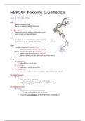
Tuesday 12th September 2023
Experiment 2: Diagnostic Restriction Digest of Human Hemoglobin Gene
Aim of the Experiment
The aim of this experiment is to investigate whether the Hemoglobin gene (HBB) is present
in a bacterial plasmid. To achieve this, the gene will be cleaved with the help of enzymes at
various positions and determined if the correct fragment sizes will appear using gel
electrophoresis.
Hypothesis
The plasmid including the HBB gene will be cleaved in 2 fragments by the BamHI enzyme.
The small fragment is expected to contain the HBB gene and have a length of 1155 base
pairs, the big fragment is expected to have a length of 9474 base pairs.
Materials and Methods
● Water
● Restriction enzymes on ice
● Enzyme buffer (cutsmart buffer)
● Heat block at 37 degrees celsius
● 6x loading dye
● Gel electrophoresis (prepared by supervisors)
● Purified plasmid, concentration approx. 150 ng / microliter
All the steps of the Protocol of experiment 1 in the Lab Manual were followed.
1. Firstly 4 µL of plasmid DNA, 2 µL of 10x cutsmart buffer, 20 µL of water were added
into one Eppendorf tube. My partner and I chose the enzyme BamHI, of which 2 µL
was added to the Eppendorf tube as well.
- The plasmid DNA, cutsmart buffer and the enzyme BamHI were pipetted with
a micropipette P20.
- The 20 µL of water was pipetted with a micropipette P200.
2. The delusion was incubated in a heat block, at 37 Celcius. This is the optimal
condition for the enzyme to exercise its function.
Due to miscommunication and time constraints, my partner and I were not able to load our
sample into the gel. We joined the group next to us, for the upcoming further steps:
This group chose BamHI, and Mfel as a second enzyme.
3. 4 µL of loading dye mix was added to the sample. This makes the sample visible and
heavier. This step was done with a micropipette P20.
4. The 20 µL sample was loaded/pipetted in one empty lane of the gel. The supervisor
added a ladder in the first lane of the gel. In this way all the known fragment lengths
would become visible.
5. The gel was run in the gel electrophoresis chamber, at 100-150V.
6. The fragment sizes of the sample were compared to the ladder.
, Results
Figure 1. The ladder used in gel electrophoresis (BMS Genetics manual page 16, 2023)
Figure 2. Obtained results gel electrophoresis
The fragment sizes that came out of the gel electrophoresis in figure 2 were interpreted
using the ladder in figure 1:
● 6000 bases and 48 ng
● 5000 bases and 40 ng