Advanced Cognitive Neurobiology and Clinical Neurophysiology
Lecture 1: Signaling within and between neurons, Bosman 2
Lecture 2: Extracellular ensemble recordings and population coding, Pennartz 6
Homework: Extracellular ensemble recordings and population coding 10
Lecture 3: Computational models I, Mejias 13
Homework: Computational models I 16
Lecture 4: Single unit recordings, Olsece 18
Lecture 5: Extracellular recordings: local field potentials, Bosman 23
Homework: Extracellular recordings: local field potentials 28
Lecture 6: Optogenetics, Olsece 32
Homework: Optogenetics 36
Lecture 7: Brain rhythms and coherence, Bosman 38
Lecture 8: Multisensory integration, Olsece 45
Homework: Multisensory integration 49
Lecture 9: Computational models II, Mejias 51
Homework: Computational models II 54
Lecture 10: Basics of consciousness, Pennartz 56
Homework: Basics of consciousness 59
Lecture 11: Human invasive electrophysiology, Bosman 61
Homework: Human invasive electrophysiology 64
Lecture 12: Mass-neural dynamics in health and disease, Linkenkaer-Hansen 67
Lecture 13: Sleep, dreaming and replay, Pennartz 68
Practicum: AMC - Pathology 72
Practicum: ratten- en schapenbrein 73
Lecture 14: Brain Computer Interfaces, Aarnoutse 74
Lecture 15: Two-photon imaging, Suzuki 76
Lecture 16: Neuropsychology of Parkinson's disease, Vriend 76
1
,Lecture 1: Signaling within and between neurons, Bosman
brain → systems → maps → networks → neurons → synapses → molecules
information by certain techniques is limited by time and space → overview of techniques is
needed to see what happens in the brain in different times and spaces
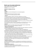
Cajal: neuron drawing, a neuron is a unity
→ information can flow between neurons
→ with this the brain can be studied as a network
timeline:
- single cell recordings were very important for Cajal in the beginning
- now we see more multicellular recordings, because we see things as a network
- 1873: Golgi method invention
- 1888: neuron doctrine
- 1929: development of EEG
Membrane potential (how does the neuron work)
- Hodgkin and Huxley
- squid, because of it’s long axons
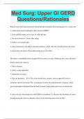
what causes a membrane potential:
1. Separation between ions in and outside the cell
2. Excess negative charge inside the cell at rest
3. The ion fluxes across the membrane
4. The lipid bilayer structure of the membrane
- Membranes are lipid bilayers
- Membrane potential arises from a separation of positive and negative charges
across the cell membrane
- Excess charges are concentrated directly at the membrane (in- and outside)
- Each excess is only a tiny fraction of the total number of ions in/outside the cell
- More potassium (K) inside the cell, more sodium (Na) outside
- negative charges inside the cell
- positive charges outside the cell
- membrane potential is used for speed in the communication; rapid transport of
signals through CNS → you only have to open the gates to use the ion gradients.
- membrane potential: Vm = Vin - Vout
- resting membrane potential: -85 mV tot -60mV
- Polarization: potential at which Vin –Vout is not zero.
- Depolarization: loss of (negative) polarization, so Vm moves towards 0 mV
- Hyperpolarization: reinforced (negative)polarization, so Vm moves further away from
0 mV, becomes more negative than before
- Convention: direction of current flow is defined as direction of net movement of
positive charge.
2
,Ion channels
Ion channels are the gateway through the membrane (because membranes are lipid
bilayers, they are not permeable to molecules or water)
- have a selectivity for size and binding site
- open and close in response to electrical, mechanical
and chemical signals
- is very rapid
- is passive
- direction and magnitude of flux are dependent on
- electrostatic forces (across membrane)
- concentration difference (in vs outside cell)
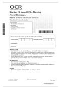
- ion channels can be:
- open
- closed
- inactive / refractory: is open, but blocked. A way to control channels
different types
1. ligand gated: sensitive to chemical substance, e.g. transmitter or olfactory stimulus
2. phosphorylation gated: ATP needed
3. voltage gated: sensitive to transmembrane voltage
4. mechanically gated: sensitive to pressure or stretch / volume change
5. resting channels: non-gated, normally open as leak current to keep the neuron at a
resting membrane potential
Equilibrium potential
Equilibrium potential: potential at which chemical driving force == electrical driving force →
no net flux of ions (Nernst equation)
Resting potential is determined by a weighted sum of the equilibrium potentials, where the
weight is determined by the relative permeability of the ion
3
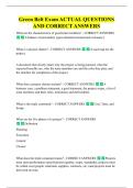
, Steady-state condition with passive current flow balanced by active ion transport by the
Na+/K+ pump.
steady-state: no net current flow into or from cell (but ATP needed to maintain) → stable
membrane potential
Local signalling
3 passive electrical properties:
1. input resistance
a. change in electric current and transmembrane voltage
2. membrane capacitance
a. Phospholipid bilayer has the ability to (transiently) “store” electrical charges
depending on membrane potential.
3. axial resistance
a. in cytoplasm
myelination: glial cell membranes are tightly wrapped around axons, the thicker the
insulation, the lower the capacitance → faster spreading to neighboring regions
node of ranvier: amount of current flowing down axon from axon hillock is not enough to
discharge capacitance along the entire length of the axon → nodes of Ranvier prevent the
action potential from dying out.
Action potentials
Time course of events:
1) Initial depolarization needed
2) Na+ channels open
3) Further depolarization of membrane
4) More Na+ channels open → more depolarization (positive feedback loop, regenerative)
5) K+ channels start to open: repolarizing effect
6) Na+ channels inactivate
7) K+ conductance outlasts Na+ conductance: hyperpolarization
8) Refractory period → impossible or very difficult to excite membrane
- two things need to happen at the same time to reach hyperpolarization;
- sodium (Na) channels close
- potassium (K) channels open
K+ conductance involved in AP:
- Grows with stronger depolarization
- Does not inactivate
4
Lecture 1: Signaling within and between neurons, Bosman 2
Lecture 2: Extracellular ensemble recordings and population coding, Pennartz 6
Homework: Extracellular ensemble recordings and population coding 10
Lecture 3: Computational models I, Mejias 13
Homework: Computational models I 16
Lecture 4: Single unit recordings, Olsece 18
Lecture 5: Extracellular recordings: local field potentials, Bosman 23
Homework: Extracellular recordings: local field potentials 28
Lecture 6: Optogenetics, Olsece 32
Homework: Optogenetics 36
Lecture 7: Brain rhythms and coherence, Bosman 38
Lecture 8: Multisensory integration, Olsece 45
Homework: Multisensory integration 49
Lecture 9: Computational models II, Mejias 51
Homework: Computational models II 54
Lecture 10: Basics of consciousness, Pennartz 56
Homework: Basics of consciousness 59
Lecture 11: Human invasive electrophysiology, Bosman 61
Homework: Human invasive electrophysiology 64
Lecture 12: Mass-neural dynamics in health and disease, Linkenkaer-Hansen 67
Lecture 13: Sleep, dreaming and replay, Pennartz 68
Practicum: AMC - Pathology 72
Practicum: ratten- en schapenbrein 73
Lecture 14: Brain Computer Interfaces, Aarnoutse 74
Lecture 15: Two-photon imaging, Suzuki 76
Lecture 16: Neuropsychology of Parkinson's disease, Vriend 76
1
,Lecture 1: Signaling within and between neurons, Bosman
brain → systems → maps → networks → neurons → synapses → molecules
information by certain techniques is limited by time and space → overview of techniques is
needed to see what happens in the brain in different times and spaces
Cajal: neuron drawing, a neuron is a unity
→ information can flow between neurons
→ with this the brain can be studied as a network
timeline:
- single cell recordings were very important for Cajal in the beginning
- now we see more multicellular recordings, because we see things as a network
- 1873: Golgi method invention
- 1888: neuron doctrine
- 1929: development of EEG
Membrane potential (how does the neuron work)
- Hodgkin and Huxley
- squid, because of it’s long axons
what causes a membrane potential:
1. Separation between ions in and outside the cell
2. Excess negative charge inside the cell at rest
3. The ion fluxes across the membrane
4. The lipid bilayer structure of the membrane
- Membranes are lipid bilayers
- Membrane potential arises from a separation of positive and negative charges
across the cell membrane
- Excess charges are concentrated directly at the membrane (in- and outside)
- Each excess is only a tiny fraction of the total number of ions in/outside the cell
- More potassium (K) inside the cell, more sodium (Na) outside
- negative charges inside the cell
- positive charges outside the cell
- membrane potential is used for speed in the communication; rapid transport of
signals through CNS → you only have to open the gates to use the ion gradients.
- membrane potential: Vm = Vin - Vout
- resting membrane potential: -85 mV tot -60mV
- Polarization: potential at which Vin –Vout is not zero.
- Depolarization: loss of (negative) polarization, so Vm moves towards 0 mV
- Hyperpolarization: reinforced (negative)polarization, so Vm moves further away from
0 mV, becomes more negative than before
- Convention: direction of current flow is defined as direction of net movement of
positive charge.
2
,Ion channels
Ion channels are the gateway through the membrane (because membranes are lipid
bilayers, they are not permeable to molecules or water)
- have a selectivity for size and binding site
- open and close in response to electrical, mechanical
and chemical signals
- is very rapid
- is passive
- direction and magnitude of flux are dependent on
- electrostatic forces (across membrane)
- concentration difference (in vs outside cell)
- ion channels can be:
- open
- closed
- inactive / refractory: is open, but blocked. A way to control channels
different types
1. ligand gated: sensitive to chemical substance, e.g. transmitter or olfactory stimulus
2. phosphorylation gated: ATP needed
3. voltage gated: sensitive to transmembrane voltage
4. mechanically gated: sensitive to pressure or stretch / volume change
5. resting channels: non-gated, normally open as leak current to keep the neuron at a
resting membrane potential
Equilibrium potential
Equilibrium potential: potential at which chemical driving force == electrical driving force →
no net flux of ions (Nernst equation)
Resting potential is determined by a weighted sum of the equilibrium potentials, where the
weight is determined by the relative permeability of the ion
3
, Steady-state condition with passive current flow balanced by active ion transport by the
Na+/K+ pump.
steady-state: no net current flow into or from cell (but ATP needed to maintain) → stable
membrane potential
Local signalling
3 passive electrical properties:
1. input resistance
a. change in electric current and transmembrane voltage
2. membrane capacitance
a. Phospholipid bilayer has the ability to (transiently) “store” electrical charges
depending on membrane potential.
3. axial resistance
a. in cytoplasm
myelination: glial cell membranes are tightly wrapped around axons, the thicker the
insulation, the lower the capacitance → faster spreading to neighboring regions
node of ranvier: amount of current flowing down axon from axon hillock is not enough to
discharge capacitance along the entire length of the axon → nodes of Ranvier prevent the
action potential from dying out.
Action potentials
Time course of events:
1) Initial depolarization needed
2) Na+ channels open
3) Further depolarization of membrane
4) More Na+ channels open → more depolarization (positive feedback loop, regenerative)
5) K+ channels start to open: repolarizing effect
6) Na+ channels inactivate
7) K+ conductance outlasts Na+ conductance: hyperpolarization
8) Refractory period → impossible or very difficult to excite membrane
- two things need to happen at the same time to reach hyperpolarization;
- sodium (Na) channels close
- potassium (K) channels open
K+ conductance involved in AP:
- Grows with stronger depolarization
- Does not inactivate
4