3.6.3 Skeletal muscles acting as effectors – AQA A Level Biology Summary Notes
3 types of muscles: skeletal (bones), smooth (small intestine, trachea and bronchioles) and cardiac
muscle (heart)
Note: skeletal muscle can also be called striated, striped or voluntary muscle
(Skeletal) Muscles act in antagonistic pairs against an incompressible skeleton
An incompresible skeleton is a skeleton that cannot be deformed by force
Antagonistic pairs refers to the fact muscles can only contract, thus for movement in two directions
muscles must work in pairs with one muscle pulling a bone in one direction and the second muscle in
the pair contracting to cause movement in the opposite direction
Muscles are used to maintain posture. This occurs when antagonistic muscles both contract at a
certain joint to keep the body at a certain angle. The contraction is isometric (doesn’t involve
movement) and only a few muscle fibres are used to avoid fatigue
Flexion = shortening
Extension = lengthening
Agonist = contracts
Antagonist = relaxes
Tendons = muscle to bone
Ligaments = bone to bone
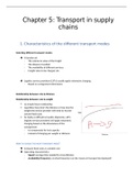
Gross and microscopic structure of skeletal muscle
Muscles have evolved by the individual cells fusing to form long muscle fibres. These muscle fibres
contain:
- Many nuclei
- Mitocondria
- Sarcolemma (cell surface membrane)
- Transverse tubules (T tubules)
- Sarcoplasm (cytoplasm)
- Sarcoplasmic reticulum that stores Ca2+
- Each individual muscle contains various proteins arranged to form myofibrils
, Sarcolemma: plasma membrane of muscle fibres
Sarcoplasm: cytoplasm of muscle fibres
The sarcolemma is folded inwards and these tubes stick into the sarcoplasm. These tubes are called transverse T
tubules.
Sarcoplasmic reticulum: endoplasmic reticulum of muscle fibres. It stores and releases Ca2+ that is needed for
muscle contraction
There are many mitochondria to provide the ATP for muscle contraction
Myofibrils are long cylindrical organelles specialised for muscle fibre contraction
3 types of muscles: skeletal (bones), smooth (small intestine, trachea and bronchioles) and cardiac
muscle (heart)
Note: skeletal muscle can also be called striated, striped or voluntary muscle
(Skeletal) Muscles act in antagonistic pairs against an incompressible skeleton
An incompresible skeleton is a skeleton that cannot be deformed by force
Antagonistic pairs refers to the fact muscles can only contract, thus for movement in two directions
muscles must work in pairs with one muscle pulling a bone in one direction and the second muscle in
the pair contracting to cause movement in the opposite direction
Muscles are used to maintain posture. This occurs when antagonistic muscles both contract at a
certain joint to keep the body at a certain angle. The contraction is isometric (doesn’t involve
movement) and only a few muscle fibres are used to avoid fatigue
Flexion = shortening
Extension = lengthening
Agonist = contracts
Antagonist = relaxes
Tendons = muscle to bone
Ligaments = bone to bone
Gross and microscopic structure of skeletal muscle
Muscles have evolved by the individual cells fusing to form long muscle fibres. These muscle fibres
contain:
- Many nuclei
- Mitocondria
- Sarcolemma (cell surface membrane)
- Transverse tubules (T tubules)
- Sarcoplasm (cytoplasm)
- Sarcoplasmic reticulum that stores Ca2+
- Each individual muscle contains various proteins arranged to form myofibrils
, Sarcolemma: plasma membrane of muscle fibres
Sarcoplasm: cytoplasm of muscle fibres
The sarcolemma is folded inwards and these tubes stick into the sarcoplasm. These tubes are called transverse T
tubules.
Sarcoplasmic reticulum: endoplasmic reticulum of muscle fibres. It stores and releases Ca2+ that is needed for
muscle contraction
There are many mitochondria to provide the ATP for muscle contraction
Myofibrils are long cylindrical organelles specialised for muscle fibre contraction