3. Functional Magnetic Resonance Imaging
1. Functional Neuroimaging: Basic Idea
o Neural activity consumes energy. To provides this energy, the cardiovascular system delivers glucose
and oxygen towards the active brain regions. Therefore, if we can track cerebral blood flow (and its
contents), we can localize regions with high activations.
o Why is localization important?
A common view of the mind (and brain) is that it is modularised, with different modules being
involved in specific functions.
The idea of modularity is around for quite a time, and although challenged more recently, still
provides a parsimonious framework for how the mind is organized
By localising different cognitive functions, we can, for example, make inferences about their role in
the processing hierarchy or their similarity or dissimilarity
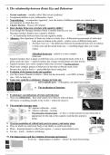
o Components of an MRI scanner- Three key components:
A big magnet, creating a strong static magnetic field (>1 Tesla)
Radiofrequency coils, to apply radiofrequency pulses to the participants
Gradient coils, modulating the magnetic field
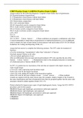
o MRI Safety - Strong magnetic fields yield hazards:
Ferromagnetic objects are very strongly attracted
Metallic objects can move or heat up
General rule #1: Do under no circumstances get close to an MRI with any metal on you!
General rule #2: The scanner is always ON!
2. Mr Physics and Structural Imaging
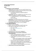
o Nuclear Spins and Precession
Atoms are composed of protons, which have different quantum spins (often described as a “turning”
around an axis > magnetic orientation).
When atoms have an odd amount of nuclear spins, atoms have a magnetic moment: They behave like
little magnetic rods. Most prominently found in the human body, these are protons (H+).
The magnetization of these nuclei is exploited in MRI (MRI was previously called “Nuclear
Magnetic Resonance”)
In addition to their magnetic moment, the protons “precess” around their magnetic moment: they
constantly wobble (invarte) around their magnetic direction.
This will become important for measuring the MRI signal later…
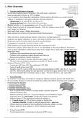
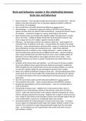
o Aligning Nuclear Spins and Net Magnetization
Normally, spin directions are randomly distributed
Applying a constant external magnetic field aligns the spins with the external field
From now on, we will look at “voxels” as spatial measurement units
The size of the voxels determines the spatial resolution of the fMRI measurements
The voxel size we can achieve depends on multiple factors, such as the
scanner used and the type of image taken
Importantly, there is an overall net magnetization: Around 6 spins per
million align along a preferential direction (at a field strength of 1Tesla)
These few per million are the signal we work with. The higher the field
strength, the higher the net magnetization, the better the signal!
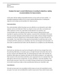
Spins forming the net magnetisation all point in the same direction, creating
a magnetisation along the direction of the external field (here z-direction),
which is usually along the body
But their precession (their “wiggling”) is not aligned, and therefore no
magnetization orthogonal to the z-direction
o Using Magnetic Resonance to Measure a Signal
Spins prefer to be aligned with the external magnetic field, as this is the
lowest-energy state
1. Functional Neuroimaging: Basic Idea
o Neural activity consumes energy. To provides this energy, the cardiovascular system delivers glucose
and oxygen towards the active brain regions. Therefore, if we can track cerebral blood flow (and its
contents), we can localize regions with high activations.
o Why is localization important?
A common view of the mind (and brain) is that it is modularised, with different modules being
involved in specific functions.
The idea of modularity is around for quite a time, and although challenged more recently, still
provides a parsimonious framework for how the mind is organized
By localising different cognitive functions, we can, for example, make inferences about their role in
the processing hierarchy or their similarity or dissimilarity
o Components of an MRI scanner- Three key components:
A big magnet, creating a strong static magnetic field (>1 Tesla)
Radiofrequency coils, to apply radiofrequency pulses to the participants
Gradient coils, modulating the magnetic field
o MRI Safety - Strong magnetic fields yield hazards:
Ferromagnetic objects are very strongly attracted
Metallic objects can move or heat up
General rule #1: Do under no circumstances get close to an MRI with any metal on you!
General rule #2: The scanner is always ON!
2. Mr Physics and Structural Imaging
o Nuclear Spins and Precession
Atoms are composed of protons, which have different quantum spins (often described as a “turning”
around an axis > magnetic orientation).
When atoms have an odd amount of nuclear spins, atoms have a magnetic moment: They behave like
little magnetic rods. Most prominently found in the human body, these are protons (H+).
The magnetization of these nuclei is exploited in MRI (MRI was previously called “Nuclear
Magnetic Resonance”)
In addition to their magnetic moment, the protons “precess” around their magnetic moment: they
constantly wobble (invarte) around their magnetic direction.
This will become important for measuring the MRI signal later…
o Aligning Nuclear Spins and Net Magnetization
Normally, spin directions are randomly distributed
Applying a constant external magnetic field aligns the spins with the external field
From now on, we will look at “voxels” as spatial measurement units
The size of the voxels determines the spatial resolution of the fMRI measurements
The voxel size we can achieve depends on multiple factors, such as the
scanner used and the type of image taken
Importantly, there is an overall net magnetization: Around 6 spins per
million align along a preferential direction (at a field strength of 1Tesla)
These few per million are the signal we work with. The higher the field
strength, the higher the net magnetization, the better the signal!
Spins forming the net magnetisation all point in the same direction, creating
a magnetisation along the direction of the external field (here z-direction),
which is usually along the body
But their precession (their “wiggling”) is not aligned, and therefore no
magnetization orthogonal to the z-direction
o Using Magnetic Resonance to Measure a Signal
Spins prefer to be aligned with the external magnetic field, as this is the
lowest-energy state