The Kidneys Regulations of Water and Inorganic Ions.
Kidneys are two bean shaped organs that lay in the posterior back of the abdominal wall, mainly located
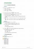
between T12, L3 VERTEBRAE, meaning they are retroperitoneal. These organs perform essential
functions in the body by filtering plasma and removing some components from the filtrate at varying
rates, depending on what the body requires.
Major functions of the Kidneys
The kidneys regulate water concentration and acid-base balance.
Regulate ions in the body.
Regulate blood pressure
Excrete waste products and foreign substances, metabolic wastes such as urea
(from breaking down of proteins), creatinine (muscle creatine), uric acid (breaking
down of nucleic acids) , toxins, drugs or hormones metabolites.
Perform Gluconeogenesis
Secrete factors and hormones into the blood such as Renin leading to the formation
of angiotensin mainly associated with blood pressure and balancing the sodium, also
produce erythropoietin and 1.25-dihydroxyvitamin D.
Regulate blood pH.
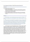
Structure of the kidneys and the urinary
The Urinary tract of a female and male.
KIDNEY
URETER
BLADDER
URETHRA
> Lay in the back of the abdominal wall
> Weighs around 150 grams
, > The urine flows from the kidneys through the ureter into the bladder, when the urinary bladder
becomes filled then the urine is emptied through the urethra, process called “micturition”.
External and Internal Anatomy of the kidney Adrenal Gland
•On top of the kidney lies the Adrenal Gland
•Has the indented area known as the hilium, consisting of the renal artery where unfiltered blood
enters and renal vein where filtered blood leaves and finally the ureter where urine passes, this region
also has lymphatics and nerves passing through.
◇Three layers of tissue surrounding the kidneys
•Surrounding innermost connective tissue, renal capsule which is tough and fibrous, protects the
kidney.
•Adipose Capsule, middle layer composed of fatty tissue, cushions the kidney from injuries.
•Renal Fascia, outer connective tissue layer.
Kidney is divided into two significant regions: Outer cortex and renal medulla
Renal medulla consists of: about 8-10 cone shaped pyramids structures arising at the dividing line
among the outer cortex and renal medulla called renal pyramids segregated by renal columns
consisting mainly of blood vessels and connective tissue, renal pyramids generate urine and is removed
into the renal papilla then emptied into ducts called minor calyx, these minor calyces join to formulate
Kidneys are two bean shaped organs that lay in the posterior back of the abdominal wall, mainly located
between T12, L3 VERTEBRAE, meaning they are retroperitoneal. These organs perform essential
functions in the body by filtering plasma and removing some components from the filtrate at varying
rates, depending on what the body requires.
Major functions of the Kidneys
The kidneys regulate water concentration and acid-base balance.
Regulate ions in the body.
Regulate blood pressure
Excrete waste products and foreign substances, metabolic wastes such as urea
(from breaking down of proteins), creatinine (muscle creatine), uric acid (breaking
down of nucleic acids) , toxins, drugs or hormones metabolites.
Perform Gluconeogenesis
Secrete factors and hormones into the blood such as Renin leading to the formation
of angiotensin mainly associated with blood pressure and balancing the sodium, also
produce erythropoietin and 1.25-dihydroxyvitamin D.
Regulate blood pH.
Structure of the kidneys and the urinary
The Urinary tract of a female and male.
KIDNEY
URETER
BLADDER
URETHRA
> Lay in the back of the abdominal wall
> Weighs around 150 grams
, > The urine flows from the kidneys through the ureter into the bladder, when the urinary bladder
becomes filled then the urine is emptied through the urethra, process called “micturition”.
External and Internal Anatomy of the kidney Adrenal Gland
•On top of the kidney lies the Adrenal Gland
•Has the indented area known as the hilium, consisting of the renal artery where unfiltered blood
enters and renal vein where filtered blood leaves and finally the ureter where urine passes, this region
also has lymphatics and nerves passing through.
◇Three layers of tissue surrounding the kidneys
•Surrounding innermost connective tissue, renal capsule which is tough and fibrous, protects the
kidney.
•Adipose Capsule, middle layer composed of fatty tissue, cushions the kidney from injuries.
•Renal Fascia, outer connective tissue layer.
Kidney is divided into two significant regions: Outer cortex and renal medulla
Renal medulla consists of: about 8-10 cone shaped pyramids structures arising at the dividing line
among the outer cortex and renal medulla called renal pyramids segregated by renal columns
consisting mainly of blood vessels and connective tissue, renal pyramids generate urine and is removed
into the renal papilla then emptied into ducts called minor calyx, these minor calyces join to formulate