MOLECULAR PRINCIPLES OF METASTASIS: A HALLMARK OF CANCER
REVIEW
Despite metastasis being the key cause of
failure of cancer therapy and mortality, it
remains poorly understood.
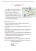
The development of metastases requires cancer
cells to leave their primary site, circulate in the
bloodstream, endure pressure in blood vessels,
acclimate to new cellular surroundings in a
secondary site, and escape deadly combat with
immune cells.
Dissemination of cancer cells precedes the
initial steps of the invasion metastasis cascade.
The cascade is the consequence of
chromosomal instability that is caused by
continuous errors in chromosome segregation
during mitosis.
In vivo and in vitro studies show that
metastatic cancer cells migrate individually. In
humans, however, it is believed that seeding
requires the joint action of a cluster of tumor
cells moving together, which bring epithelial-mesenchymal transition (EMT) into the picture.
Epithelial-Mesenchymal Transition Tumor cells that express a
Is the transdifferentiation process through which transformed epithelial cells mix of epithelial and
develop the ability to invade, resist stress, and disseminate. mesenchymal phenotypes
EMT governs the reversible biochemical alterations that permit a specific are more effective in
epithelial cell to attain a mesenchymal phenotype and confers epithelial- circulation, colonization at
mesenchymal plasticity upon epithelial cells, which is crucial for cancer the secondary site and the
progression and metastasis. development of metastasis.
According to a study of mice models, asparagine synthetase (a metabolic enzyme)
is correlated with metastasis development.
EMT program is a spectrum of transitional stages between the epithelial and
mesenchymal phenotypes. The transition of one state to another is governed by a number of growth
factors and signalling pathways.
Metastatic cells with the most pronounced mesenchymal
phenotype proliferate near endothelial and inflammatory
cells. These tumor cells release larger quantities of
chemokines and proteins to attract immune cells and
stimulate angiogenesis, thus promoting the development of a
unique inflammatory and highly vascularized niche.
Metabolic stressors, and matrix stiffness trigger the EMT
program in cancer cells.
Although EMT might be required for metastasis initiation,
the opposite process of mesenchymal- epithelial transition
(MET) is needed for metastatic progression.
Genetic Profile of Metastatic Cells
* Hundreds of genes have been reported to determine
invasive potential, suggesting that primary tumor cells
exhibit a metastatic genetic signature.
, * Most predominant genes that were somatically changed in metastasis included:
- Tumor protein p5
- Cyclin dependent kinase inhibitor 2A (CDKN2A)
- Phosphatase and tension homolog (PTEN)
- Retinoblastoma (RB1)
* Markers that predict metastatic progression showed that advanced cancers arise from diverse cell types
which deeply affects that eventual genetic and epigenetic alterations that promote metastatic
progression.
* Understanding intertumoral heterogeneity among different cancers can reveal the mechanisms of
metastatic progression and how the cell type of origin contributes to tumor development.
* The cytotoxic immune signature and the presence of lymphatic vessels play an important role in the
generation of distant metastases, regardless of genomic instability.
Metabolic Profile of Metastatic Cells
* Hypoxia-inducible factors (HIF) permit cancer cells to adapt to their cellular environment by
regulating angiogenesis, EMT, invasion, metastasis, and energy metabolism.
* HIF signalling drives the secretion of lysyl oxidase (LOX), LOX-like proteins, and exosomes, to
establish a prometastatic environment within the lung and bones of patients with breast cancer.
* Tumors with more extensive hypoxic and anoxic areas exhibit higher rates of metastasis.
* Metastatic cancer cells depend on mono-carboxylate transporter 1 (MCT1) to deal with oxidative
stress. MCT1 plays a major role in circulating lactate, which is a prominent energy source for
metastasizing cells. Highly metastatic cells have increased levels of MCT1, whereas the inhibition of
MCT1 decreases lactate uptake by metastatic cells and, thus, reduces their metastatic capability.
* Changes in ATP/ADP and ATP/AMP ratios also promote metastatic behaviour.
Priming The Premetastatic Niche
o Secondary sites do not receive invading cancer cells passively. The host microenvironment, termed the
premetastatic niche (PMN), is selectively primed by the primary tumor even before the initiation of
metastasis.
o The development of a PMN is a multistep process involving secretory factors and extracellular
vesicles that induce vascular leakage, ECM remodelling, and immunosuppression.
o Cancer cells release vesicles that carry messenger RNA transcribed from genes that are involved in
cell migration and metastasis, which are then accepted by other cells. After host cells engulf these
vesicles, human cells that did not express a malignant phenotype start to migrate faster. The
transferred genes also enhance the ability of cells to invade other organs.
o Primary tumors release significant amounts of exosomes that transfer invasion-promoting factors, such
as microRNAs (miRNAs), to tumorigenic cancer cells.
o Exosomes secrete EMT inducers that stimulate EMT progression in host epithelial cells, providing
them with the ability to invade and metastasize.
o Exosomes can remodel the ECM by interacting with fibroblasts, stromal cells, and endothelial cells to
degrade protease-associated components such as collagen, laminin, and fibronectin.
o In addition to their role in priming the PMN, exosomes exhibit properties that drive cancer cell
organotropism. This metastatic bias towards certain organs stems from exosomal avidity for specific
host cells.
o Other membrane proteins and lipids that are associated with ECM properties and adhesion influence
the specific targeting of exosomes to their specific host cells.
o In addition, exosomal internalization by target host cells activates heterogeneous endocytic pathways
such as clathrin, lipid raft, and caveolinmediated uptake.
Can Metastasis Be Driven By Epigenetic Factors?
Alterations in the motility of immune cells lead to changes in the immune microenvironment.
REVIEW
Despite metastasis being the key cause of
failure of cancer therapy and mortality, it
remains poorly understood.
The development of metastases requires cancer
cells to leave their primary site, circulate in the
bloodstream, endure pressure in blood vessels,
acclimate to new cellular surroundings in a
secondary site, and escape deadly combat with
immune cells.
Dissemination of cancer cells precedes the
initial steps of the invasion metastasis cascade.
The cascade is the consequence of
chromosomal instability that is caused by
continuous errors in chromosome segregation
during mitosis.
In vivo and in vitro studies show that
metastatic cancer cells migrate individually. In
humans, however, it is believed that seeding
requires the joint action of a cluster of tumor
cells moving together, which bring epithelial-mesenchymal transition (EMT) into the picture.
Epithelial-Mesenchymal Transition Tumor cells that express a
Is the transdifferentiation process through which transformed epithelial cells mix of epithelial and
develop the ability to invade, resist stress, and disseminate. mesenchymal phenotypes
EMT governs the reversible biochemical alterations that permit a specific are more effective in
epithelial cell to attain a mesenchymal phenotype and confers epithelial- circulation, colonization at
mesenchymal plasticity upon epithelial cells, which is crucial for cancer the secondary site and the
progression and metastasis. development of metastasis.
According to a study of mice models, asparagine synthetase (a metabolic enzyme)
is correlated with metastasis development.
EMT program is a spectrum of transitional stages between the epithelial and
mesenchymal phenotypes. The transition of one state to another is governed by a number of growth
factors and signalling pathways.
Metastatic cells with the most pronounced mesenchymal
phenotype proliferate near endothelial and inflammatory
cells. These tumor cells release larger quantities of
chemokines and proteins to attract immune cells and
stimulate angiogenesis, thus promoting the development of a
unique inflammatory and highly vascularized niche.
Metabolic stressors, and matrix stiffness trigger the EMT
program in cancer cells.
Although EMT might be required for metastasis initiation,
the opposite process of mesenchymal- epithelial transition
(MET) is needed for metastatic progression.
Genetic Profile of Metastatic Cells
* Hundreds of genes have been reported to determine
invasive potential, suggesting that primary tumor cells
exhibit a metastatic genetic signature.
, * Most predominant genes that were somatically changed in metastasis included:
- Tumor protein p5
- Cyclin dependent kinase inhibitor 2A (CDKN2A)
- Phosphatase and tension homolog (PTEN)
- Retinoblastoma (RB1)
* Markers that predict metastatic progression showed that advanced cancers arise from diverse cell types
which deeply affects that eventual genetic and epigenetic alterations that promote metastatic
progression.
* Understanding intertumoral heterogeneity among different cancers can reveal the mechanisms of
metastatic progression and how the cell type of origin contributes to tumor development.
* The cytotoxic immune signature and the presence of lymphatic vessels play an important role in the
generation of distant metastases, regardless of genomic instability.
Metabolic Profile of Metastatic Cells
* Hypoxia-inducible factors (HIF) permit cancer cells to adapt to their cellular environment by
regulating angiogenesis, EMT, invasion, metastasis, and energy metabolism.
* HIF signalling drives the secretion of lysyl oxidase (LOX), LOX-like proteins, and exosomes, to
establish a prometastatic environment within the lung and bones of patients with breast cancer.
* Tumors with more extensive hypoxic and anoxic areas exhibit higher rates of metastasis.
* Metastatic cancer cells depend on mono-carboxylate transporter 1 (MCT1) to deal with oxidative
stress. MCT1 plays a major role in circulating lactate, which is a prominent energy source for
metastasizing cells. Highly metastatic cells have increased levels of MCT1, whereas the inhibition of
MCT1 decreases lactate uptake by metastatic cells and, thus, reduces their metastatic capability.
* Changes in ATP/ADP and ATP/AMP ratios also promote metastatic behaviour.
Priming The Premetastatic Niche
o Secondary sites do not receive invading cancer cells passively. The host microenvironment, termed the
premetastatic niche (PMN), is selectively primed by the primary tumor even before the initiation of
metastasis.
o The development of a PMN is a multistep process involving secretory factors and extracellular
vesicles that induce vascular leakage, ECM remodelling, and immunosuppression.
o Cancer cells release vesicles that carry messenger RNA transcribed from genes that are involved in
cell migration and metastasis, which are then accepted by other cells. After host cells engulf these
vesicles, human cells that did not express a malignant phenotype start to migrate faster. The
transferred genes also enhance the ability of cells to invade other organs.
o Primary tumors release significant amounts of exosomes that transfer invasion-promoting factors, such
as microRNAs (miRNAs), to tumorigenic cancer cells.
o Exosomes secrete EMT inducers that stimulate EMT progression in host epithelial cells, providing
them with the ability to invade and metastasize.
o Exosomes can remodel the ECM by interacting with fibroblasts, stromal cells, and endothelial cells to
degrade protease-associated components such as collagen, laminin, and fibronectin.
o In addition to their role in priming the PMN, exosomes exhibit properties that drive cancer cell
organotropism. This metastatic bias towards certain organs stems from exosomal avidity for specific
host cells.
o Other membrane proteins and lipids that are associated with ECM properties and adhesion influence
the specific targeting of exosomes to their specific host cells.
o In addition, exosomal internalization by target host cells activates heterogeneous endocytic pathways
such as clathrin, lipid raft, and caveolinmediated uptake.
Can Metastasis Be Driven By Epigenetic Factors?
Alterations in the motility of immune cells lead to changes in the immune microenvironment.