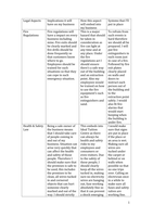
Lecture 1 – The Retina
Something from outside is projected by the lens to the retina. “This is high school stuff; I
suppose you know that”. The retina is a pre-processing unit.
The retina is a piece of neural tissue that is ‘sent out’ of the brain: it pre-processes the images
captured by the cones and rods and sends the resulting signal via the bipolar cells to the retinal
ganglion cells (RGC), then to the brain through the optic nerve.
Rods and cones are sensitive to different wavelengths:
rods are not involved in color vision but are most sensitive
to medium wavelengths.
cones are divided into three types. Blue cones best respond
to short wavelength ; green cones best respond to
medium wavelength ; red cones best respond to
long wavelength .
Photoreceptors transform light into neural signals through
proteins called rhodopsin or photopsin, for rods and cones
respectively.
I will just mention rhodopsin here, although the same applies to photopsin.
1
,Rhodopsin is activated by light, which causes its shape to change a bit, thereby activating other
proteins that cause a ‘signal cascade’, thus making the Na+ channels close.
In other words, rhodopsin translates light into the closing of Na+ channels, which causes the
membrane to hyperpolarize.
There are different versions of these opsins, which are sensitive to different wavelengths. Many
varieties can be found in the animal kingdom, where humans have 4 (3 for cones, 1 for rods).
Goldfish have 5, so they are often used in retina research.
e.g., mice have 2, which makes them less able to discriminate among different colors.
The absence of a certain type of cone causes retinal color blindness. This is
mainly studied with Ishihara plates and is 10 times less likely for females to be
color blind (it is a sex-linked genetic trait).
Photoreceptors are unevenly distributed in the retina: cones are mostly packed in the fovea
whereas rods are mostly present in the parafoveal regions.
The fovea (or macula) is the middle part of the retina: it is cup shaped and is where you have
sharp and color vision (photopic vision).
A fundoscopy checks the fundus of your eye. This
procedure shows how the light has to pass thru a lot of
obstacles to reach the photoreceptors. In fact, our retina
is covered by blood vessels and there may be ‘bugs’
floating in our vitreous humor (glassy filling of most of
our eye). These bugs are actually proteins and stuff.
The blood vessels normally receive light from the
direction of the pupil, although you can visualize them
by illuminating the side of the head, which then creates
a shadow of these blood vessels onto your retina (you
notice that they are much in the way of your retina).
Also, the fovea doesn’t have big blood vessels covering
it, which also allows a sharper vision (less obstacles).
2
,DISEASE OF THE EYE #1 (FOVEA)
• Macular Degeneration
It is caused by the accumulation of yellow deposits (dry) or blood (wet) in the macula.
These toxic products lead to photoreceptor loss.
It usually happens with age, although it is influenced by genes and diet.
It causes loss of foveal vision (blank spots), acuity loss, and distortion in central vision.
There is no treatment yet, although stem cells may be a possibility.
Besides having to pass through all the shit between the cornea and the retina, the light has to pass
through the retinal network itself in order to reach the photoreceptors.
“Now you may wonder why this is, since it looks like a fairly stupid arrangement”
The reason is that in the utmost back you have the retinal pigment epithelium (RPE): a layer of
pigment epithelium which absorbs the light, preventing it to bounce back to the photoreceptors
(which would cause the light signal to activate photoreceptors all over the place). So, the RPE
allows a ray of light to activate only a particular photoreceptor (which we need when e.g., seeing
a small detail), thus enabling really sharp vision. Finally, the RPE also provides nutrients to the
photoreceptors.
Some animals (e.g., cats) don’t have the dark RPE but have one that
reflects the light, which is why when you shine a light into their
eyes you see it reflected back to you. The advantage in this is that
the same ray of light can activate multiple photoreceptors, allowing
you to see better with low light conditions (in fact cats see better
when it is dark). However, a disadvantage is that your vision is
much less sharp (in fact, cats can see stuff that moves but don’t
have a very detailed vision of the world that surrounds them).
3
, At some point in the retina there is a blind spot (optic disk), where there are no photoreceptors
because the RGC nerve fibers pass through the optic disk and reach the brain through the optic
nerve. The optic disk is also the place where the blood vessels originate.
DISEASE OF THE EYE #2
• Glaucoma
It is caused by an increase in intraocular pressure, that is the pressure
inside the eye. In fact, the eye has to be kept at a certain pressure otherwise
it is just an empty sack. The mechanism responsible for pressure
regulation is the trabecular meshwork: if this fucks up (either by letting
the ocular fluid flow out or by not producing it anymore) the intraocular
pressure increases, causing the optic nerve to be compressed and you lose
part of your VF.
Glaucoma implies progressive loss of vision, with peripheral vision usually being the first
to degrade (because of the anatomy of the optic disk),
Figure D is full blown glaucoma: the white spot (center of
VF) is the only thing that is left.
If it is not that advanced, you may not even notice it (since
we also don’t notice our blind spot). So, it often happens
that patients notice it only when it is too late.
The treatment is to reduce pressure in your eye, which is accomplished through eyedrops
or surgery (although the already lost RGC fibers are gone).
When you are over the age of 40 or so it’s good to have your eye pressure checked. Heartfelt advice by
Victor.
You also have an acute version of glaucoma, where you get a huge increase in pressure
over a few days causing your eye to become all red and swollen, and you don’t see
anything anymore.
Glaucoma is an insidious disease, since you do not feel the increased pressure nor the loss
of peripheral vision, unless it is too widespread aka too late.
4