NURSING 3313 Exam 5
Review-pharm
Questions and Answers
• Hypothalamus and
pituitary
o Hypothalamus secretes releasing hormones to the pituitary
o Pituitary hormones go to specific tissues
o For instance:
▪ Hypothalamus secretes thyrotropin-releasing hormone (TRH)
…telling the pituitary to secrete thyroid stimulating hormone
(TSH)…TSH is secreted and acts on the thyroid to stimulate
thyroid hormone secretion
o The hypothalamus is the coordinating center for the nervous and
endocrine responses to internal and external stimuli. The
hypothalamus constantly monitors the body’s homeostasis by
analyzing input from the periphery and the central nervous system
(CNS) and coordinating responses through the autonomic, endocrine,
and nervous systems. In effect, it is the “master gland” of the
neuroendocrine system. This title was once given to the pituitary
gland because of its many functions and well-protected location.
o The hypothalamus has various regions or clusters of neurons that are
sensitive to certain stimuli. It is responsible for regulating a number
of body functions, including body temperature, thirst, hunger, water
retention, blood pressure, respiration, reproduction, and emotional
reactions. Situated at the base of the forebrain, the hypothalamus
receives input from virtually all other areas of the brain, including
the limbic system, cerebral cortex, and the special senses that are
controlled by the cranial nerves—smell, sight, touch, taste, and
hearing. Because of its positioning, the hypothalamus is able to
influence and be influenced by emotions and thoughts. The
hypothalamus is also located in an area of the brain that is poorly
protected by the blood–brain barrier, so it is able to act as a sensor
to various electrolytes, chemicals, and hormones that are in
circulation and do not affect other areas of the brain.
o The hypothalamus maintains internal homeostasis by sensing blood
chemistries and by stimulating or suppressing endocrine, autonomic,
,and CNS activity. In essence, it can turn the autonomic nervous
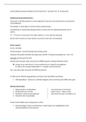
system and its effects on or off. The hypothalamus also produces and
secretes a number of releasing hormones or factors that stimulate
the pituitary gland, which in turn stimulates or inhibits various
endocrine glands throughout the body (Fig. 34.1). These releasing
hormones include growth hormone (GH)-releasing hormone,
thyrotropin-releasing hormone (TRH), gonadotropin-releasing
hormone, corticotropin-releasing hormone, and prolactin-releasing
hormone. The hypothalamus also produces two inhibiting factors
that act as regulators to shut off the production of hormones when
levels become too high: GH release–inhibiting factor (somatostatin)
and prolactin (PRL)-inhibiting factor (PIF). Recent research has
indicated that PIF may actually be dopamine, a neurotransmitter.
Patients who are taking dopamine-blocking drugs often develop
galactorrhea (inappropriate milk production) and breast
enlargement, theoretically because PIF is also blocked and PRL levels
continue to rise, stimulating breast tissue and milk production.
Research is ongoing about the chemical structure of several of the
releasing factors.
,o The hypothalamus is connected to the pituitary gland by two
networks: a vascular capillary network carries the hypothalamic-
releasing factors directly into the anterior pituitary and a
neurological network delivers two other hypothalamic hormones—
antidiuretic hormone (ADH) and oxytocin—to the posterior pituitary
to be stored. These hormones are released as needed by the body
when stimulated by the hypothalamus.
o As the “master gland” of the neuroendocrine system, the
hypothalamus helps regulate the central and autonomic nervous
systems and the endocrine system to maintain homeostasis.
o The hypothalamus produces stimulating and inhibiting factors that
travel to the anterior pituitary through a capillary system to
stimulate the release of pituitary hormones or block the production
of certain pituitary hormones when levels of target hormones get
too high.
o The hypothalamus is connected to the posterior pituitary by a
nerve network that delivers the hypothalamic hormones ADH
and oxytocin to be stored in the posterior pituitary until the
hypothalamus stimulates their release.
o Because of its position in the brain, the hypothalamus is stimulated
by many things, such as light, emotion, cerebral cortex activity, and a
variety of chemical and hormonal stimuli. Together, the
hypothalamus and the pituitary function closely to maintain
endocrine activity along what is called the hypothalamic–pituitary
axis (HPA) using a series of negative feedback systems.
o It is thought that this feedback system is more complex than once
believed. The hypothalamus probably also senses TRH and TSH
levels and regulates TRH secretion within a narrow range, even if
thyroid hormone is not produced. The anterior pituitary may also
be sensitive to TSH levels and thyroid hormone, regulating its own
production of TSH. This complex system provides backup controls
and regulation if any part of the HPA fails. This system also can
create complications, especially when there is a need to override or
interact with the total system, as is the case with hormone
replacement therapy or the treatment of endocrine disorders.
Supplying an exogenous hormone, for example, may increase the
, hormone levels in the body but then may affect the HPA to stop
production of releasing and stimulating hormones, leading to a
decrease in the body’s normal production of the hormone.
o Two of the anterior pituitary hormones (i.e., GH and PRL) do not
have a target organ to produce hormones and so cannot be
regulated by the same type of feedback mechanism. The
hypothalamus in this case responds directly to rising levels of GH and
PRL. When levels rise, the hypothalamus releases the inhibiting
factors somatostatin and PIF directly to inhibit the pituitary’s release
of GH and PRL, respectively. The HPA functions through negative
feedback loops or the direct use of inhibiting factors to constantly
keep these hormones regulated.
o
o The pituitary gland is located in the skull in the bony sella turcica
under a layer of dura mater. It is divided into three lobes: an
anterior lobe, a posterior lobe, and an
Review-pharm
Questions and Answers
• Hypothalamus and
pituitary
o Hypothalamus secretes releasing hormones to the pituitary
o Pituitary hormones go to specific tissues
o For instance:
▪ Hypothalamus secretes thyrotropin-releasing hormone (TRH)
…telling the pituitary to secrete thyroid stimulating hormone
(TSH)…TSH is secreted and acts on the thyroid to stimulate
thyroid hormone secretion
o The hypothalamus is the coordinating center for the nervous and
endocrine responses to internal and external stimuli. The
hypothalamus constantly monitors the body’s homeostasis by
analyzing input from the periphery and the central nervous system
(CNS) and coordinating responses through the autonomic, endocrine,
and nervous systems. In effect, it is the “master gland” of the
neuroendocrine system. This title was once given to the pituitary
gland because of its many functions and well-protected location.
o The hypothalamus has various regions or clusters of neurons that are
sensitive to certain stimuli. It is responsible for regulating a number
of body functions, including body temperature, thirst, hunger, water
retention, blood pressure, respiration, reproduction, and emotional
reactions. Situated at the base of the forebrain, the hypothalamus
receives input from virtually all other areas of the brain, including
the limbic system, cerebral cortex, and the special senses that are
controlled by the cranial nerves—smell, sight, touch, taste, and
hearing. Because of its positioning, the hypothalamus is able to
influence and be influenced by emotions and thoughts. The
hypothalamus is also located in an area of the brain that is poorly
protected by the blood–brain barrier, so it is able to act as a sensor
to various electrolytes, chemicals, and hormones that are in
circulation and do not affect other areas of the brain.
o The hypothalamus maintains internal homeostasis by sensing blood
chemistries and by stimulating or suppressing endocrine, autonomic,
,and CNS activity. In essence, it can turn the autonomic nervous
system and its effects on or off. The hypothalamus also produces and
secretes a number of releasing hormones or factors that stimulate
the pituitary gland, which in turn stimulates or inhibits various
endocrine glands throughout the body (Fig. 34.1). These releasing
hormones include growth hormone (GH)-releasing hormone,
thyrotropin-releasing hormone (TRH), gonadotropin-releasing
hormone, corticotropin-releasing hormone, and prolactin-releasing
hormone. The hypothalamus also produces two inhibiting factors
that act as regulators to shut off the production of hormones when
levels become too high: GH release–inhibiting factor (somatostatin)
and prolactin (PRL)-inhibiting factor (PIF). Recent research has
indicated that PIF may actually be dopamine, a neurotransmitter.
Patients who are taking dopamine-blocking drugs often develop
galactorrhea (inappropriate milk production) and breast
enlargement, theoretically because PIF is also blocked and PRL levels
continue to rise, stimulating breast tissue and milk production.
Research is ongoing about the chemical structure of several of the
releasing factors.
,o The hypothalamus is connected to the pituitary gland by two
networks: a vascular capillary network carries the hypothalamic-
releasing factors directly into the anterior pituitary and a
neurological network delivers two other hypothalamic hormones—
antidiuretic hormone (ADH) and oxytocin—to the posterior pituitary
to be stored. These hormones are released as needed by the body
when stimulated by the hypothalamus.
o As the “master gland” of the neuroendocrine system, the
hypothalamus helps regulate the central and autonomic nervous
systems and the endocrine system to maintain homeostasis.
o The hypothalamus produces stimulating and inhibiting factors that
travel to the anterior pituitary through a capillary system to
stimulate the release of pituitary hormones or block the production
of certain pituitary hormones when levels of target hormones get
too high.
o The hypothalamus is connected to the posterior pituitary by a
nerve network that delivers the hypothalamic hormones ADH
and oxytocin to be stored in the posterior pituitary until the
hypothalamus stimulates their release.
o Because of its position in the brain, the hypothalamus is stimulated
by many things, such as light, emotion, cerebral cortex activity, and a
variety of chemical and hormonal stimuli. Together, the
hypothalamus and the pituitary function closely to maintain
endocrine activity along what is called the hypothalamic–pituitary
axis (HPA) using a series of negative feedback systems.
o It is thought that this feedback system is more complex than once
believed. The hypothalamus probably also senses TRH and TSH
levels and regulates TRH secretion within a narrow range, even if
thyroid hormone is not produced. The anterior pituitary may also
be sensitive to TSH levels and thyroid hormone, regulating its own
production of TSH. This complex system provides backup controls
and regulation if any part of the HPA fails. This system also can
create complications, especially when there is a need to override or
interact with the total system, as is the case with hormone
replacement therapy or the treatment of endocrine disorders.
Supplying an exogenous hormone, for example, may increase the
, hormone levels in the body but then may affect the HPA to stop
production of releasing and stimulating hormones, leading to a
decrease in the body’s normal production of the hormone.
o Two of the anterior pituitary hormones (i.e., GH and PRL) do not
have a target organ to produce hormones and so cannot be
regulated by the same type of feedback mechanism. The
hypothalamus in this case responds directly to rising levels of GH and
PRL. When levels rise, the hypothalamus releases the inhibiting
factors somatostatin and PIF directly to inhibit the pituitary’s release
of GH and PRL, respectively. The HPA functions through negative
feedback loops or the direct use of inhibiting factors to constantly
keep these hormones regulated.
o
o The pituitary gland is located in the skull in the bony sella turcica
under a layer of dura mater. It is divided into three lobes: an
anterior lobe, a posterior lobe, and an