IIF Revision Notes
[Topic 6] Immunity, Infection and Forensics
, A Level Notes IIF: Splicing & DNA Profiling Biology
Transcription:
1) RNA polymerase attaches to DNA double helix at start codon (beginning of gene)
2) Hydrogen bonds break, separating strands and DNA unwinds/uncoils
3) Antisense strand (template) used to make mRNA copy
4) RNA polymerase lines up free floating mononucleotides; complementary base pairing (U instead of T) – mRNA
creates reverse copy of antisense strand
5) Phosphodiester bonds link RNA nucleotides; chain separates as mRNA molecule
6) Hydrogen bonds reform; coil into double helix
7) RNA polymerase reaches stop codon and detaches from DNA
8) mRNA leaves via nuclear pore and attaches to ribosome where translation occurs
Translation:
1) mRNA attaches to ribosome and tRNA carry amino acids to ribosome
2) tRNA has anticodon complementary to mRNA start codon; attaches via complementary base pairing
3) Second tRNA attaches to next condon in same way
4) Two amino acids attached to tRNA molecules are joined by a peptide bond; tRNA leaves
5) Ribosome moves along to next codon; process repeats until mRNA stop codon
6) Polypeptide chain produced moves away from ribosome and translation complete
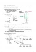
Splicing is the cleaving/attachment/joining of exons subsequent to removal/discarding of introns.
After transcription has occurred, pre-mRNA is produced; tRNA has anticodons CCA, GAA
• exons (expressed segment of gene) Complementary codons GGU, CUU
Complementary triplet code CCA, GAA
• introns (intermediate non-coding segment of gene)
Alternative RNA splicing occurs whereby introns are removed and exons are joined together; this mature mRNA that is
produced has undergone post-transcriptional change which can then be translated.
During RNA splicing different exon are removed which results in more than one mature mRNA strands arising from one
gene; hence one gene does not code for one protein.
When an intron is accidently kept in the mature mRNA an error has occurred; many cancers result from mutant
proteins being produced when introns are retained.
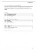
DNA profiling is used to determine genetic relationships between organisms.
1) DNA sample obtained from muscle, saliva, semen or blood
2) Restriction enzymes (cut out STR) applied, cutting DNA into fragments; sample amplified by PCR
3) Add dye/fluorescent tag
4) Use pipette to place sample on gel
5) Apply electrical current; small fragments faster than larger ones; UV light reveals positions
6) This is gel electrophoresis (produces bands of STR fragments)
✓ Bands in the same position contain similar fragments
✓ The more similar patterns are, the closer the relationship
✓ If testing a different species, it shows they shared a recent common ancestor
Restriction enzymes are used to cut DNA at precise points; they cut at a
recognition site which consists of a particular sequence of bases – they are also
called endonucleases because they cut the DNA at sites within the DNA strand.
When restriction enzymes cut DNA they can leave a blunt end or sticky ends
(e.g. 4 base pair sticky end); this means it cuts at one point and ends cutting 4
base pair later at a point same as the first (e.g. start and end at C|G).
[Topic 6] Immunity, Infection and Forensics
, A Level Notes IIF: Splicing & DNA Profiling Biology
Transcription:
1) RNA polymerase attaches to DNA double helix at start codon (beginning of gene)
2) Hydrogen bonds break, separating strands and DNA unwinds/uncoils
3) Antisense strand (template) used to make mRNA copy
4) RNA polymerase lines up free floating mononucleotides; complementary base pairing (U instead of T) – mRNA
creates reverse copy of antisense strand
5) Phosphodiester bonds link RNA nucleotides; chain separates as mRNA molecule
6) Hydrogen bonds reform; coil into double helix
7) RNA polymerase reaches stop codon and detaches from DNA
8) mRNA leaves via nuclear pore and attaches to ribosome where translation occurs
Translation:
1) mRNA attaches to ribosome and tRNA carry amino acids to ribosome
2) tRNA has anticodon complementary to mRNA start codon; attaches via complementary base pairing
3) Second tRNA attaches to next condon in same way
4) Two amino acids attached to tRNA molecules are joined by a peptide bond; tRNA leaves
5) Ribosome moves along to next codon; process repeats until mRNA stop codon
6) Polypeptide chain produced moves away from ribosome and translation complete
Splicing is the cleaving/attachment/joining of exons subsequent to removal/discarding of introns.
After transcription has occurred, pre-mRNA is produced; tRNA has anticodons CCA, GAA
• exons (expressed segment of gene) Complementary codons GGU, CUU
Complementary triplet code CCA, GAA
• introns (intermediate non-coding segment of gene)
Alternative RNA splicing occurs whereby introns are removed and exons are joined together; this mature mRNA that is
produced has undergone post-transcriptional change which can then be translated.
During RNA splicing different exon are removed which results in more than one mature mRNA strands arising from one
gene; hence one gene does not code for one protein.
When an intron is accidently kept in the mature mRNA an error has occurred; many cancers result from mutant
proteins being produced when introns are retained.
DNA profiling is used to determine genetic relationships between organisms.
1) DNA sample obtained from muscle, saliva, semen or blood
2) Restriction enzymes (cut out STR) applied, cutting DNA into fragments; sample amplified by PCR
3) Add dye/fluorescent tag
4) Use pipette to place sample on gel
5) Apply electrical current; small fragments faster than larger ones; UV light reveals positions
6) This is gel electrophoresis (produces bands of STR fragments)
✓ Bands in the same position contain similar fragments
✓ The more similar patterns are, the closer the relationship
✓ If testing a different species, it shows they shared a recent common ancestor
Restriction enzymes are used to cut DNA at precise points; they cut at a
recognition site which consists of a particular sequence of bases – they are also
called endonucleases because they cut the DNA at sites within the DNA strand.
When restriction enzymes cut DNA they can leave a blunt end or sticky ends
(e.g. 4 base pair sticky end); this means it cuts at one point and ends cutting 4
base pair later at a point same as the first (e.g. start and end at C|G).