Table of Contents
Musculoskeletal System ......................................................................................... 1
Lecture 2 – Limb Development ......................................................................................... 1
Lecture 3 – Joint Structure & Function ............................................................................. 4
Lecture 4 – Muscle Disorders ........................................................................................... 7
Lecture 5 - Pain ............................................................................................................... 10
Lecture 6 – Musculoskeletal Pain ................................................................................... 13
Lecture 7 – Bone & Cartilage .......................................................................................... 15
Lecture 8 – Inflammatory Arthritis ................................................................................. 19
Lecture 9 – Rheumatoid Arthritis ................................................................................... 22
Lecture 10 – Osteoporosis .............................................................................................. 24
Lectures 11 & 12– Back Pain I & II (Physiotherapy and Surgery) .................................... 27
Lecture 13 – Ageing of the Musculoskeletal System ...................................................... 28
Lectures 14 & 15 – Osteoarthritis ................................................................................... 29
Lecture 16 – Drug Mechanisms of Action for RA, OA and Gout ..................................... 33
Musculoskeletal System
Lecture 2 – Limb Development
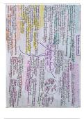
• How do our growth plates shut down when we are done growing? The fibroblast growth
factor receptor-3 (FGFR-3) is activated by fibroblast growth factor (FGF), which closes the
growth plates and stops up from growing anymore
• What is achondroplasia and what’s it caused by? It’s one of the most common causes of
dwarfism. In achondroplasia, the growth plates close early because there is a mutation in
the FGFR-3 that makes it activated even when there is no FGF about. This gives rise to
people with short limbs (because the growth plates closed early) but a normal sized torso
and head.
• How have we learnt most of what we know about limb development? Cutting a bit of limb
bud and see how it affects the resulting chick embryo
• How long does it take for your whole skeleton to be laid down in a mini-cartilage
framework form during embryonic development? By the end of the first trimester
• In what order do you lay down the cartilage framework of the limbs? Proximal to distal, for
example you’d lay down the framework for your humerus, before your
radius/ulna, before your phalanges.
• What evidence is there that they are laid down proximal à distal? If
you trim off the very tip of the limb bud (apical ectodermal ridge) as it’s
developing, you end up with a limb that is underdeveloped. If you trim
the tip off early, you end up with only a humerus, trim it a little bit later
and you end up with half a radius and ulna as well, and if you trim it
even later you have a whole arm that is missing the fingertips…and so
on.
• What is the structure of this special ‘lip’ that seems to be controlling
growth? It’s pseudostratified epithelium, as opposed to the rest of the
embryo which is mostly simple epithelium. These cells release a growth
Simple epithelium
The tip that can be trimmed in
the above experiment, which is
known as the apical ectodermal
ridge (AER)
, signal that tells the developing mesenchymal cells what to do.
• How does the apical ectodermal ridge control the growth of the mesenchymal cells?
o The AER (and only the AER) releases a signal, indicated by the arrows
o This signal can only diffuse a certain distance, and the area within this distance
which is bathed in signal is called the progress zone
o Any cell in the progress zone is close enough to the AER to ‘smell’ the signal
o So long as the cell can ‘smell’ the signal, it carries on dividing
o Eventually, the cell gets pushed out of the progress zone by newly divided cells, at
which point the cell stops going through cell divisions
o The cell somehow knows how many cell divisions it has gone through in total
o If the cell has gone through only a few cell divisions, it will become a proximal
structure, for example a humerus. If the cell has gone through many cell divisions, it
will become a distal structure, for example a phalanx
o It may be helpful to think of this process using a ‘clock’ metaphor:
Ø The AER (and only the AER) releases a signal, indicated by the arrows
Ø This signal can only diffuse a certain distance, and the area within this distance
which is bathed in signal is called the progress zone
Ø Any cell in the progress zone is close enough to
the AER to ‘smell’ the signal
Ø The signal makes their innate ‘clock’ keep
ticking forward
Ø So long as the cell can ‘smell’ the signal, it’s
clock keeps ticking forward
Ø As soon as they are pushed out of the progress
zone by newly divided cells, their clock stops
Ø The ‘time’ on the ‘clock’ determines what
structure that cell will become along the
proximal-distal axis.
Ø If the clock stops early, it’ll be a proximal structure, and if the clock stops late
then it’ll be a distal structure
Ø The ‘clock’ is used as a metaphor for number of cell divisions
2
,• What is the name of this signal made by the AER? Its called ridge factor, but it’s actually
FGF, fibroblast growth factor (the same growth factor that tells you to close your growth
plates)
• How do the cells know how many cell cycles they’ve been through? Using
‘HOX genes’.
• What was the thalidomide disaster? A morning sickness pill that lead to
countless deformities in the babies, mainly with limb development that
often lead to children having their hands poking out of their shoulders with
no humerus, radius or ulna, and equivalent in the lower limb sometimes.
• What went wrong during limb development of thalidomide? Some of the 0
cells in the diagram above were killed due to the thalidomide which killed the vessels
supplying this area. This meant that surviving cells could divide, staying in the progress zone,
without being forced out by dividing cells, because most of them had been killed. This leads
to a lack of cells that have undergone 1 or 2 cell divisions (and hence would have ended up
as humerus, radius/ulna), and only cells that have undergone 3 divisions. This explains why
you get distal structures (hands) poking out of the torso, as opposed to a smooth
progression of proximal à distal structures
• So far we have explained proximal à distal development (shoulder à hand), but how do
we get anterio-posterior development (head à tail or thumb à pinky)?
o The posterior side (pinky) has high ZPA = zona polarising activity due to the presence
of a ZPA factor
o The ZPA factor diffuses anteriorly towards the thumb, but when it gets there it’s
obviously at a lower concentration
o [ZPA factor] = anterio-posterior placement, so if you’re a cell with a lot of ZPA factor,
you’ll be a posterior structure (pinky), and if there’s not a lot of ZPA factor, you’ll be
an anterior structure (thumb)
• What is the evidence for this? If you graft the posterior side of the hand of a donor chick
embryo onto the anterior side of a recipient chick embryo, you end up with a mirror image
hand with two thumbs in the middle (anterior in the middle) and pinky’s on the outside
(posterior on either side)
• What are hedgehog genes? Genes involved in anterio-posterior development
• What is the most important one in humans? A gene called sonic hedgehog
• What does the sonic hedgehog protein do? It diffuses a very short distance (not nearly
enough to get to the anterior side from the posterior side), activating another gene called
the BMP gene (bimorph genetic protein gene).
• What does the BMP gene do? It codes for BMP, which is the ZPA factor in humans. A high
[BMP] à posterior structure (pinky or ulna)…. A low [BMP] à anterior structure (thumb or
radius)
3
, • Overall, what tells you what structure to be in the limb? Its like an x,y coordinate plot
where a high [FGF] tells you to be a proximal structure and a high [BMP] tells you to be a
posterior structure. For example:
Structure [FGF] [BMP]
Humerus High -
Radius Medium Low
Ulna Medium High
Thumb Low Low
Pinky Low High
• What happens if you graft a bit of a leg bud onto an arm
bud just beneath the apical ectodermal ridge? The cells
still somehow know they are meant to be leg tissue, but
their ‘clock’ is restarted so the number of cell divisions
restarts meaning a bit of tissue that was going to end up
femur, if placed under the AER of the wing bud, will turn
into toes on the end of a wing.
• So how do cells know whether to be an arm or a leg?
Thanks to special T-box genes that are turned on before
any proximal-distal patterning occurs. So arm knows it’ll
be arm before it knows whether it will be a humerus of a
finger tip
• How do muscles and nerves develop over the cartilage
framework? Whatever type of connective tissue/cartilage
(i.e. arm, leg, forearm) is there, the surrounding
mesenchymal cells will develop according to that. The connective tissue is the boss and tells
the muscle how to grow. Once the muscles and skin have grown, this tells the nerves how to
grow (hierarchy = connective tissue à muscle à nerve)
• How do joints form? Due to the movement of skeletal elements during development and
due to genes such as GDF5, BMP2 and NOG.
Lecture 3 – Joint Structure & Function
• What are the two ways to classify joints and what are their subtypes?
1. According to degree of motion
o Synarthroses: no/little movement
o Amphiarthrosis: some movement
o Diarthroses: free movement
2. According to structure
o Fibrous (3 types)
i. Sutures – found in the skull and can either
be serrated, lap or plane sutures depending
on how the two bones fit together (see
diagram)
ii. Syndesmosis – when bones are joined by an
intraosseous ligament or dense aponeurotic
membrane
iii. Gomphosis – a peg and socket joint held
together by collagenous connective tissue,
found between the teeth and the jaw
(which have periodontal ligaments)
o Cartilaginous (2 types)
4
Musculoskeletal System ......................................................................................... 1
Lecture 2 – Limb Development ......................................................................................... 1
Lecture 3 – Joint Structure & Function ............................................................................. 4
Lecture 4 – Muscle Disorders ........................................................................................... 7
Lecture 5 - Pain ............................................................................................................... 10
Lecture 6 – Musculoskeletal Pain ................................................................................... 13
Lecture 7 – Bone & Cartilage .......................................................................................... 15
Lecture 8 – Inflammatory Arthritis ................................................................................. 19
Lecture 9 – Rheumatoid Arthritis ................................................................................... 22
Lecture 10 – Osteoporosis .............................................................................................. 24
Lectures 11 & 12– Back Pain I & II (Physiotherapy and Surgery) .................................... 27
Lecture 13 – Ageing of the Musculoskeletal System ...................................................... 28
Lectures 14 & 15 – Osteoarthritis ................................................................................... 29
Lecture 16 – Drug Mechanisms of Action for RA, OA and Gout ..................................... 33
Musculoskeletal System
Lecture 2 – Limb Development
• How do our growth plates shut down when we are done growing? The fibroblast growth
factor receptor-3 (FGFR-3) is activated by fibroblast growth factor (FGF), which closes the
growth plates and stops up from growing anymore
• What is achondroplasia and what’s it caused by? It’s one of the most common causes of
dwarfism. In achondroplasia, the growth plates close early because there is a mutation in
the FGFR-3 that makes it activated even when there is no FGF about. This gives rise to
people with short limbs (because the growth plates closed early) but a normal sized torso
and head.
• How have we learnt most of what we know about limb development? Cutting a bit of limb
bud and see how it affects the resulting chick embryo
• How long does it take for your whole skeleton to be laid down in a mini-cartilage
framework form during embryonic development? By the end of the first trimester
• In what order do you lay down the cartilage framework of the limbs? Proximal to distal, for
example you’d lay down the framework for your humerus, before your
radius/ulna, before your phalanges.
• What evidence is there that they are laid down proximal à distal? If
you trim off the very tip of the limb bud (apical ectodermal ridge) as it’s
developing, you end up with a limb that is underdeveloped. If you trim
the tip off early, you end up with only a humerus, trim it a little bit later
and you end up with half a radius and ulna as well, and if you trim it
even later you have a whole arm that is missing the fingertips…and so
on.
• What is the structure of this special ‘lip’ that seems to be controlling
growth? It’s pseudostratified epithelium, as opposed to the rest of the
embryo which is mostly simple epithelium. These cells release a growth
Simple epithelium
The tip that can be trimmed in
the above experiment, which is
known as the apical ectodermal
ridge (AER)
, signal that tells the developing mesenchymal cells what to do.
• How does the apical ectodermal ridge control the growth of the mesenchymal cells?
o The AER (and only the AER) releases a signal, indicated by the arrows
o This signal can only diffuse a certain distance, and the area within this distance
which is bathed in signal is called the progress zone
o Any cell in the progress zone is close enough to the AER to ‘smell’ the signal
o So long as the cell can ‘smell’ the signal, it carries on dividing
o Eventually, the cell gets pushed out of the progress zone by newly divided cells, at
which point the cell stops going through cell divisions
o The cell somehow knows how many cell divisions it has gone through in total
o If the cell has gone through only a few cell divisions, it will become a proximal
structure, for example a humerus. If the cell has gone through many cell divisions, it
will become a distal structure, for example a phalanx
o It may be helpful to think of this process using a ‘clock’ metaphor:
Ø The AER (and only the AER) releases a signal, indicated by the arrows
Ø This signal can only diffuse a certain distance, and the area within this distance
which is bathed in signal is called the progress zone
Ø Any cell in the progress zone is close enough to
the AER to ‘smell’ the signal
Ø The signal makes their innate ‘clock’ keep
ticking forward
Ø So long as the cell can ‘smell’ the signal, it’s
clock keeps ticking forward
Ø As soon as they are pushed out of the progress
zone by newly divided cells, their clock stops
Ø The ‘time’ on the ‘clock’ determines what
structure that cell will become along the
proximal-distal axis.
Ø If the clock stops early, it’ll be a proximal structure, and if the clock stops late
then it’ll be a distal structure
Ø The ‘clock’ is used as a metaphor for number of cell divisions
2
,• What is the name of this signal made by the AER? Its called ridge factor, but it’s actually
FGF, fibroblast growth factor (the same growth factor that tells you to close your growth
plates)
• How do the cells know how many cell cycles they’ve been through? Using
‘HOX genes’.
• What was the thalidomide disaster? A morning sickness pill that lead to
countless deformities in the babies, mainly with limb development that
often lead to children having their hands poking out of their shoulders with
no humerus, radius or ulna, and equivalent in the lower limb sometimes.
• What went wrong during limb development of thalidomide? Some of the 0
cells in the diagram above were killed due to the thalidomide which killed the vessels
supplying this area. This meant that surviving cells could divide, staying in the progress zone,
without being forced out by dividing cells, because most of them had been killed. This leads
to a lack of cells that have undergone 1 or 2 cell divisions (and hence would have ended up
as humerus, radius/ulna), and only cells that have undergone 3 divisions. This explains why
you get distal structures (hands) poking out of the torso, as opposed to a smooth
progression of proximal à distal structures
• So far we have explained proximal à distal development (shoulder à hand), but how do
we get anterio-posterior development (head à tail or thumb à pinky)?
o The posterior side (pinky) has high ZPA = zona polarising activity due to the presence
of a ZPA factor
o The ZPA factor diffuses anteriorly towards the thumb, but when it gets there it’s
obviously at a lower concentration
o [ZPA factor] = anterio-posterior placement, so if you’re a cell with a lot of ZPA factor,
you’ll be a posterior structure (pinky), and if there’s not a lot of ZPA factor, you’ll be
an anterior structure (thumb)
• What is the evidence for this? If you graft the posterior side of the hand of a donor chick
embryo onto the anterior side of a recipient chick embryo, you end up with a mirror image
hand with two thumbs in the middle (anterior in the middle) and pinky’s on the outside
(posterior on either side)
• What are hedgehog genes? Genes involved in anterio-posterior development
• What is the most important one in humans? A gene called sonic hedgehog
• What does the sonic hedgehog protein do? It diffuses a very short distance (not nearly
enough to get to the anterior side from the posterior side), activating another gene called
the BMP gene (bimorph genetic protein gene).
• What does the BMP gene do? It codes for BMP, which is the ZPA factor in humans. A high
[BMP] à posterior structure (pinky or ulna)…. A low [BMP] à anterior structure (thumb or
radius)
3
, • Overall, what tells you what structure to be in the limb? Its like an x,y coordinate plot
where a high [FGF] tells you to be a proximal structure and a high [BMP] tells you to be a
posterior structure. For example:
Structure [FGF] [BMP]
Humerus High -
Radius Medium Low
Ulna Medium High
Thumb Low Low
Pinky Low High
• What happens if you graft a bit of a leg bud onto an arm
bud just beneath the apical ectodermal ridge? The cells
still somehow know they are meant to be leg tissue, but
their ‘clock’ is restarted so the number of cell divisions
restarts meaning a bit of tissue that was going to end up
femur, if placed under the AER of the wing bud, will turn
into toes on the end of a wing.
• So how do cells know whether to be an arm or a leg?
Thanks to special T-box genes that are turned on before
any proximal-distal patterning occurs. So arm knows it’ll
be arm before it knows whether it will be a humerus of a
finger tip
• How do muscles and nerves develop over the cartilage
framework? Whatever type of connective tissue/cartilage
(i.e. arm, leg, forearm) is there, the surrounding
mesenchymal cells will develop according to that. The connective tissue is the boss and tells
the muscle how to grow. Once the muscles and skin have grown, this tells the nerves how to
grow (hierarchy = connective tissue à muscle à nerve)
• How do joints form? Due to the movement of skeletal elements during development and
due to genes such as GDF5, BMP2 and NOG.
Lecture 3 – Joint Structure & Function
• What are the two ways to classify joints and what are their subtypes?
1. According to degree of motion
o Synarthroses: no/little movement
o Amphiarthrosis: some movement
o Diarthroses: free movement
2. According to structure
o Fibrous (3 types)
i. Sutures – found in the skull and can either
be serrated, lap or plane sutures depending
on how the two bones fit together (see
diagram)
ii. Syndesmosis – when bones are joined by an
intraosseous ligament or dense aponeurotic
membrane
iii. Gomphosis – a peg and socket joint held
together by collagenous connective tissue,
found between the teeth and the jaw
(which have periodontal ligaments)
o Cartilaginous (2 types)
4