Ziekteleer onderste lidmaat: HEUP
Pijnlijke heup bij volwassene
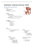
a. Heup
= coxofemoraal gewricht
= kogelgewricht
Bal (femurkop) in kom (acetabulum)
Acetabulum
o Hyalien kraakbeen
o Rondom labrum (270°)
Zorgt voor extra stabiliteit zodat het beter past
o Lig. transversum
o Enkel perifeer gedeelte = bedekt met kraakbeen
Heup mobiliteit
o Kogelgewricht rotatie
Flexie: 120°
Extensie: 30°
Exorotatie: 30-45°
Endorotatie: 20-35°
Adductie: 20-30°
Abductie: 45°
Spieren
o Flexoren
M. iliopsoas (belangrijkst! Filet pur) o Extensoren
M. rectus femoris M. gluteus maximus
M. sartorius (belangrijkst)
Hamstrings
M. biceps femoris
M. semitendinosus
M. semimembranosus
o Adductoren
M. gluteus medius o Abductoren (indien geïnhibeert:
M. gluteus minimus Trendelenburg)
Adductoren
M. adductor magnus M. gluteus medius
M. adductor longus M. gluteus minimus
M. adductor brevis
(Vaak eerst aangetast)
, o Exorotatoren
M. piriformis o Endorotatoren
M. obturatorius internus en externus Geen echte endorotator
Mm. Gemelli Meerdere spieren als
M. quadratus femoris secundaire actie
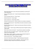
Krachtverdeling heup bij monopodaal steunen (tredelenburg)
o Om lichaam in evenwicht te houden adductoren 3 x grotere spierkracht > gewicht
o A = normaal
Bekken horizontaal
o B = positief
Bekken kantelen
o Door pathologie in de heup zijn de heupabductoren dermate geïnhibeerd, zodat het
bekken kantelt waarbij de contralaterale zijde naar onderen zakt
,Klinisch onderzoek
o Intra articulair heup:
Pijn in lies
Soms uitstralingen naar knie C-sign
o Pijn t.h.v. trochanter:
Meestal tendinitis (peesontsteking) of bursitis (ontstoken slijmbeurs)
o Pijn t.h.v. bil:
Eerder onderrug
o Gangpatroon:
Verkorte steun
Trendelenburg
Externe rotatiegang
Voet naar buiten om heup te ontspannen
o Verminderde interne rotatie
= 1 van de éérste tekenen van een aandoening in de heup
Test van Patrick
o Diagnose voor waar de pijn vandaan komt
o = Faber test
Flexie, Abductie, Externe Rotatie
o Been in cijfer 4 positie, knie naar beneden duwen
o Pijn posterieur of bil sacro-iliacaal
o Pijn in lies iliopsoas-pees
Test van Thomas
o Voor patiënten met heupflexiecontracturen
o Gezond been in hyperflexie brengen
Verhinderd compensaties van bekken
o Positief been kan niet blijven liggen op onderzoekstafel
Aangetaste heup gaat in flexie
flexiecontractuur
, b. Coxartrose (artrosis deformans)
- Slijtage van kraakbeen
Reactieve inflammatie (ontsteking) van het synovium
- Vorming osteofyten (botuitsteeksels of papegaaienbekken) en gewrichtsmuizen
- oorzaken
o Primair (idiopatisch): meest frequent
Door slijtage, ouderdom, pure pech
o Secundair
Door trauma
Inflammatoire (reumatische) arthritis
Avasculaire necrose
Afsterven door doorbloedingsstoornissen
Sekwel aandoening heup tijdens jeugd
Legg-Calve-Perthes (4-10j) => bloedvoorzieningsstoornis door
groeien, waardoor heup kop vervormd
epifysiolyse (10-15j) => afschuiven kop door wegvallen epifysaire
schijf
Vormafwijking: dysplasie
- Symptomen
o Pijn tijdens/na belasten
o Minder pijn bij rust
o Pijnpunten:
Lies
Voorzijde dij – knie
Grote trochanter
Gluteaalstreek
o Ochtendstijfheid en startpijn
Meestal minder dan 30 min.
Indien langer eerder reumatisch
o Naarmate artrose zich meer inzet nachtelijke pijn en rustpijn
o Bewegingsbeperking:
Moeilijkheden om schoen of kousen aan of uit te trekken
o Manken
- Diagnose
o Staande radiografie
Staand omdat zo een gewrichtsspleetvernauwing meer opvalt
Osteofyten
Sclerose van het bot verharding van het bot
Bot ziet er witter uit
Subchondrale cysten - geoden (= kleine openingen die zich vormen onder het
kraakbeen)
Pijnlijke heup bij volwassene
a. Heup
= coxofemoraal gewricht
= kogelgewricht
Bal (femurkop) in kom (acetabulum)
Acetabulum
o Hyalien kraakbeen
o Rondom labrum (270°)
Zorgt voor extra stabiliteit zodat het beter past
o Lig. transversum
o Enkel perifeer gedeelte = bedekt met kraakbeen
Heup mobiliteit
o Kogelgewricht rotatie
Flexie: 120°
Extensie: 30°
Exorotatie: 30-45°
Endorotatie: 20-35°
Adductie: 20-30°
Abductie: 45°
Spieren
o Flexoren
M. iliopsoas (belangrijkst! Filet pur) o Extensoren
M. rectus femoris M. gluteus maximus
M. sartorius (belangrijkst)
Hamstrings
M. biceps femoris
M. semitendinosus
M. semimembranosus
o Adductoren
M. gluteus medius o Abductoren (indien geïnhibeert:
M. gluteus minimus Trendelenburg)
Adductoren
M. adductor magnus M. gluteus medius
M. adductor longus M. gluteus minimus
M. adductor brevis
(Vaak eerst aangetast)
, o Exorotatoren
M. piriformis o Endorotatoren
M. obturatorius internus en externus Geen echte endorotator
Mm. Gemelli Meerdere spieren als
M. quadratus femoris secundaire actie
Krachtverdeling heup bij monopodaal steunen (tredelenburg)
o Om lichaam in evenwicht te houden adductoren 3 x grotere spierkracht > gewicht
o A = normaal
Bekken horizontaal
o B = positief
Bekken kantelen
o Door pathologie in de heup zijn de heupabductoren dermate geïnhibeerd, zodat het
bekken kantelt waarbij de contralaterale zijde naar onderen zakt
,Klinisch onderzoek
o Intra articulair heup:
Pijn in lies
Soms uitstralingen naar knie C-sign
o Pijn t.h.v. trochanter:
Meestal tendinitis (peesontsteking) of bursitis (ontstoken slijmbeurs)
o Pijn t.h.v. bil:
Eerder onderrug
o Gangpatroon:
Verkorte steun
Trendelenburg
Externe rotatiegang
Voet naar buiten om heup te ontspannen
o Verminderde interne rotatie
= 1 van de éérste tekenen van een aandoening in de heup
Test van Patrick
o Diagnose voor waar de pijn vandaan komt
o = Faber test
Flexie, Abductie, Externe Rotatie
o Been in cijfer 4 positie, knie naar beneden duwen
o Pijn posterieur of bil sacro-iliacaal
o Pijn in lies iliopsoas-pees
Test van Thomas
o Voor patiënten met heupflexiecontracturen
o Gezond been in hyperflexie brengen
Verhinderd compensaties van bekken
o Positief been kan niet blijven liggen op onderzoekstafel
Aangetaste heup gaat in flexie
flexiecontractuur
, b. Coxartrose (artrosis deformans)
- Slijtage van kraakbeen
Reactieve inflammatie (ontsteking) van het synovium
- Vorming osteofyten (botuitsteeksels of papegaaienbekken) en gewrichtsmuizen
- oorzaken
o Primair (idiopatisch): meest frequent
Door slijtage, ouderdom, pure pech
o Secundair
Door trauma
Inflammatoire (reumatische) arthritis
Avasculaire necrose
Afsterven door doorbloedingsstoornissen
Sekwel aandoening heup tijdens jeugd
Legg-Calve-Perthes (4-10j) => bloedvoorzieningsstoornis door
groeien, waardoor heup kop vervormd
epifysiolyse (10-15j) => afschuiven kop door wegvallen epifysaire
schijf
Vormafwijking: dysplasie
- Symptomen
o Pijn tijdens/na belasten
o Minder pijn bij rust
o Pijnpunten:
Lies
Voorzijde dij – knie
Grote trochanter
Gluteaalstreek
o Ochtendstijfheid en startpijn
Meestal minder dan 30 min.
Indien langer eerder reumatisch
o Naarmate artrose zich meer inzet nachtelijke pijn en rustpijn
o Bewegingsbeperking:
Moeilijkheden om schoen of kousen aan of uit te trekken
o Manken
- Diagnose
o Staande radiografie
Staand omdat zo een gewrichtsspleetvernauwing meer opvalt
Osteofyten
Sclerose van het bot verharding van het bot
Bot ziet er witter uit
Subchondrale cysten - geoden (= kleine openingen die zich vormen onder het
kraakbeen)