1.4 SUMMARY
PROBLEM 1
BIOLOGICAL VOCAB
Dorsal: toward the back, ventral: toward the stomach.
Horizontal: parallel to the horizon, sagittal: divides the body into
right and left, frontal: divides the body into dorsal and ventral.
Anterior: toward to front end (göğüs), posterior: toward the rear
end (sırt)
Lateral: towards side, away from midline, medial: towards
midline, away from side.
Ipsilateral: on the same side of the body, contralateral: on the
opposite side of body.
Clusters of cell bodies in CNS is nuclei, clusters of cell bodies in
PNS is ganglia (daha küçük ama sayıca daha fazla)
Bundles of axons in CNS is tracts, bundles of axons in PNS is
nerves.
NEURONS
1) Axons: Myelin sheath (fatty insulation around axon) covers, myelin
sheaths protect the axon and impulse. Have specific gaps between
myelin sheaths called nodes of ranvier. They pass information to
long distances and have axon branches which passes the
information to different directions.
2) Dendrites: collection of information from other neurons, they have
branches with fibers, they have synaptic receptors. (The grater the
surface area of dendrites, more information they can receive.
3) Soma/cell body: Includes nucleus and other organelles. Location of
DNA (cell nucleus)
4) Presynaptic terminals/terminal buttons: Send neurotransmitters to
the synaptic cleft, connection with other neurons.
Neurons do not have centrosomes so they can’t reproduce.
Neurons have lot of mitochondria bc they require energy for
impulse transformation.
1) Sensory neurons (afferent axon-brings in the info-): Neurons
responsible for converting external stimuli from the environment to
an internal stimuli. They feel sensory output via their receptors.
(Sense external stimuli)
2) Motor neurons (efferent axon -carries info away): Mostly due
within muscles or glands. They respond to the CNS.
3) Inter neurons: Communication between sensory, motor neurons
that are in CNS. No or small axon. Integration of neural activity no
to pass on information.
When sensory neurons are damaged: no feeling is felt. (local
anesthesia)
Inter neurons are damaged: people feel things, unable to interpret
them. (paralysis)
Motor neurons are damaged: feeling is felt, interpretion have been
made but one can’t control their muscles. (Pain is felt but they can’t
go away from the pain)
Neurons cell membrane: lipid bilayer- selectively permeable.
, A neuron with more than 2 processes emanating from their cell
bodies are multipolar, only 1 process is unipolar, and 2 processes
is bipolar neuron.
GLIAL CELLS
Remove waste material as viruses, fungi or other microorganisms,
helps neurons, support and feed neurons. Come glial cells form
myelin sheaths.
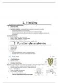
BRAIN
Consists of 3 parts. (Forebrain, midbrain and
hindbrain).
Gryus or gyri (çıkıntı), sulcus (girinti), sulci
(pl)
Forebrain
Consists of 2 cerebral hemispheres. (right and
left)
Outer portion is the cerebral cortex.
Telencephalon (cerebral cortex, limbic system, basal ganglia, cerebral
commissures)
CEREBRAL CORTEX (lobes, major fissures and gyri)
Consists of 4 lobes.
Frontal lobe
Controls primary motor cortex (precentral gyrus: location of
primary motor cortex) (control of fine movement)
Problem solving, thinking, planning.
Prefrontal cortex: the very anterior portion of frontal lobe.
(organizing behavior)
Temporal lobe
Cortical target for auditory info.
Left temporal lobe: understanding spoken language.
Contains hippocampus.
Hallucinations due to tumors.
Parietal Lobe
Has somatosensory cortex (postcentral gyrus: location of
somatosensory cortex): receives sensory information from touch
receptors and muscle stretch receptors.
Pressure, light-touch information.
Orientation, movement, recognition.
Occipital lobe
Visual information.
Contains primary visual cortex in the posterior pole.
Damage: cortical blindness.
Eyes provide stimuli, visual cortex provides experience. (?)
Central fissure/girinti
Separates frontal and parietal lobe. (primary sensory cortex:
parietal lobe, primary motor cortex: frontal lobe)
Lateral fissure
Separates the temporal lobe from the frontal and parietal lobes.
Longitudinal fissure/sulcus
Deep grove that separates two hemispheres.
,Precentral gyrus
Site of primary motor cortex.
Postcentral gyrus
Location of primary somatosensory cortex.
Superior temporal gyrus
Contains auditory cortex.
Cingulate gyrus
Curved fold around the corpus callosum. (component of limbic
system)
LIMBIC SYSTEM (regulates motivated behavior)
Amygdala
Respond to emotional information. (mostly fear) (4F’s: fighting,
fleeing, feeding and fornicating) (Located in temporal lobe)
Hippocampus
Long term memory, orientation, face and language recognition.
(temporal lobe)
Fornix
Connects hippocampus to the rest of the brain. (C SHAPE)
Cingulate cortex
Learning, emotions and memory. (located in cingulate gyrus)
Septum
Separates hemispheres (in the middle)
BASAL GANGLIA (learning, emotions)
Striatum
Caudate nucleus
Control of movement, decision making. (damage: Alzheimer,
Parkinson disease)
Putamen
Globus pallidus
Nucleus accumbens
Reward system, dopamine.
CEREBRAL COMMISSURES
Corpus callossum
Connection between hemispheres.
Anterior commissures, posterior commissures
Other connections between hemispheres
Diencephalon
Thalamus
Receives sensory information and sends it to cerebral cortex.
Olfactory information does not go to thalamus.
Integration of information.
Hypothalamus
Voluntary actions (eating, drinking, sleeping, mating)
Responsible for homeostasis.
Conveys messages to pituitary gland.
Arcuate nucleus, interstitial nucleus, suprachiasmatic nucleus,
paraventricular nucleus, lateral nucleus.
Optic chiasm
, The point where the optic nerves from each eye come together.
Visual field
Pituitary gland
Secrete hormones according to hypothalamus
Pineal gland
Nucleus basalis
Midbrain (Mesencephalon)
Tectum
Root of the midbrain.
Has superior colliculus (vision) and inferior colliculus (hearing).
(Both are important for sensory processing)
Tegmentum
Under tectum.
Substantia nigra, red nucleus: responsible for movement.
(dopamine). (sensory motor system)
Ventral tegmental area: reward system, motivation, cognition and
drug addiction.
Cerebral aqueduct: connects the 3rd and 4th ventricles
Reticular formation: reflexes, consciousness, alertness.
Periaqueductual gray: Suppresses pain
Hindbrain
Consists of medulla, pons and cerebellum.
Medulla, pons and midbrain forms brainstem. (beyin sapı)
Reticular formation.
Medulla (medulla oblongata) (omurilik soğanı) (Myelencephalon)
Controls vital reflexes through cranial nerves. (breathing, heart
rate, vomiting, salivation, coughing.)
Damage to medulla is fatal.
Non-voluntary functions.
Pons (Metencephalon)
Contains nuclei for several cranial nerves.
Axons from each half of brain is crossed to the opposite
side of the spinal cord, so left hemisphere controls the
muscles on the right side and opposite.
Reticular formation (motor and sensory nerves pass)
Plays role in sleep and arousal (locus coeruleus)
Cerebellum (Beyincik) (Metencephalon)
Control of movement.
Balance and coordination.
Motor memory.
The Ventricles
4 fluid filled cavities called the ventricles.
Ventricles have cells called choroid plexus which produce
cerebrospinal fluid (CSF) a clear fluid similar to blood plasma.
Ventricles shock absorption, protector.
CSF fills the ventricles and the meninges membrane that surround
the brain and the spinal cord.
PROBLEM 1
BIOLOGICAL VOCAB
Dorsal: toward the back, ventral: toward the stomach.
Horizontal: parallel to the horizon, sagittal: divides the body into
right and left, frontal: divides the body into dorsal and ventral.
Anterior: toward to front end (göğüs), posterior: toward the rear
end (sırt)
Lateral: towards side, away from midline, medial: towards
midline, away from side.
Ipsilateral: on the same side of the body, contralateral: on the
opposite side of body.
Clusters of cell bodies in CNS is nuclei, clusters of cell bodies in
PNS is ganglia (daha küçük ama sayıca daha fazla)
Bundles of axons in CNS is tracts, bundles of axons in PNS is
nerves.
NEURONS
1) Axons: Myelin sheath (fatty insulation around axon) covers, myelin
sheaths protect the axon and impulse. Have specific gaps between
myelin sheaths called nodes of ranvier. They pass information to
long distances and have axon branches which passes the
information to different directions.
2) Dendrites: collection of information from other neurons, they have
branches with fibers, they have synaptic receptors. (The grater the
surface area of dendrites, more information they can receive.
3) Soma/cell body: Includes nucleus and other organelles. Location of
DNA (cell nucleus)
4) Presynaptic terminals/terminal buttons: Send neurotransmitters to
the synaptic cleft, connection with other neurons.
Neurons do not have centrosomes so they can’t reproduce.
Neurons have lot of mitochondria bc they require energy for
impulse transformation.
1) Sensory neurons (afferent axon-brings in the info-): Neurons
responsible for converting external stimuli from the environment to
an internal stimuli. They feel sensory output via their receptors.
(Sense external stimuli)
2) Motor neurons (efferent axon -carries info away): Mostly due
within muscles or glands. They respond to the CNS.
3) Inter neurons: Communication between sensory, motor neurons
that are in CNS. No or small axon. Integration of neural activity no
to pass on information.
When sensory neurons are damaged: no feeling is felt. (local
anesthesia)
Inter neurons are damaged: people feel things, unable to interpret
them. (paralysis)
Motor neurons are damaged: feeling is felt, interpretion have been
made but one can’t control their muscles. (Pain is felt but they can’t
go away from the pain)
Neurons cell membrane: lipid bilayer- selectively permeable.
, A neuron with more than 2 processes emanating from their cell
bodies are multipolar, only 1 process is unipolar, and 2 processes
is bipolar neuron.
GLIAL CELLS
Remove waste material as viruses, fungi or other microorganisms,
helps neurons, support and feed neurons. Come glial cells form
myelin sheaths.
BRAIN
Consists of 3 parts. (Forebrain, midbrain and
hindbrain).
Gryus or gyri (çıkıntı), sulcus (girinti), sulci
(pl)
Forebrain
Consists of 2 cerebral hemispheres. (right and
left)
Outer portion is the cerebral cortex.
Telencephalon (cerebral cortex, limbic system, basal ganglia, cerebral
commissures)
CEREBRAL CORTEX (lobes, major fissures and gyri)
Consists of 4 lobes.
Frontal lobe
Controls primary motor cortex (precentral gyrus: location of
primary motor cortex) (control of fine movement)
Problem solving, thinking, planning.
Prefrontal cortex: the very anterior portion of frontal lobe.
(organizing behavior)
Temporal lobe
Cortical target for auditory info.
Left temporal lobe: understanding spoken language.
Contains hippocampus.
Hallucinations due to tumors.
Parietal Lobe
Has somatosensory cortex (postcentral gyrus: location of
somatosensory cortex): receives sensory information from touch
receptors and muscle stretch receptors.
Pressure, light-touch information.
Orientation, movement, recognition.
Occipital lobe
Visual information.
Contains primary visual cortex in the posterior pole.
Damage: cortical blindness.
Eyes provide stimuli, visual cortex provides experience. (?)
Central fissure/girinti
Separates frontal and parietal lobe. (primary sensory cortex:
parietal lobe, primary motor cortex: frontal lobe)
Lateral fissure
Separates the temporal lobe from the frontal and parietal lobes.
Longitudinal fissure/sulcus
Deep grove that separates two hemispheres.
,Precentral gyrus
Site of primary motor cortex.
Postcentral gyrus
Location of primary somatosensory cortex.
Superior temporal gyrus
Contains auditory cortex.
Cingulate gyrus
Curved fold around the corpus callosum. (component of limbic
system)
LIMBIC SYSTEM (regulates motivated behavior)
Amygdala
Respond to emotional information. (mostly fear) (4F’s: fighting,
fleeing, feeding and fornicating) (Located in temporal lobe)
Hippocampus
Long term memory, orientation, face and language recognition.
(temporal lobe)
Fornix
Connects hippocampus to the rest of the brain. (C SHAPE)
Cingulate cortex
Learning, emotions and memory. (located in cingulate gyrus)
Septum
Separates hemispheres (in the middle)
BASAL GANGLIA (learning, emotions)
Striatum
Caudate nucleus
Control of movement, decision making. (damage: Alzheimer,
Parkinson disease)
Putamen
Globus pallidus
Nucleus accumbens
Reward system, dopamine.
CEREBRAL COMMISSURES
Corpus callossum
Connection between hemispheres.
Anterior commissures, posterior commissures
Other connections between hemispheres
Diencephalon
Thalamus
Receives sensory information and sends it to cerebral cortex.
Olfactory information does not go to thalamus.
Integration of information.
Hypothalamus
Voluntary actions (eating, drinking, sleeping, mating)
Responsible for homeostasis.
Conveys messages to pituitary gland.
Arcuate nucleus, interstitial nucleus, suprachiasmatic nucleus,
paraventricular nucleus, lateral nucleus.
Optic chiasm
, The point where the optic nerves from each eye come together.
Visual field
Pituitary gland
Secrete hormones according to hypothalamus
Pineal gland
Nucleus basalis
Midbrain (Mesencephalon)
Tectum
Root of the midbrain.
Has superior colliculus (vision) and inferior colliculus (hearing).
(Both are important for sensory processing)
Tegmentum
Under tectum.
Substantia nigra, red nucleus: responsible for movement.
(dopamine). (sensory motor system)
Ventral tegmental area: reward system, motivation, cognition and
drug addiction.
Cerebral aqueduct: connects the 3rd and 4th ventricles
Reticular formation: reflexes, consciousness, alertness.
Periaqueductual gray: Suppresses pain
Hindbrain
Consists of medulla, pons and cerebellum.
Medulla, pons and midbrain forms brainstem. (beyin sapı)
Reticular formation.
Medulla (medulla oblongata) (omurilik soğanı) (Myelencephalon)
Controls vital reflexes through cranial nerves. (breathing, heart
rate, vomiting, salivation, coughing.)
Damage to medulla is fatal.
Non-voluntary functions.
Pons (Metencephalon)
Contains nuclei for several cranial nerves.
Axons from each half of brain is crossed to the opposite
side of the spinal cord, so left hemisphere controls the
muscles on the right side and opposite.
Reticular formation (motor and sensory nerves pass)
Plays role in sleep and arousal (locus coeruleus)
Cerebellum (Beyincik) (Metencephalon)
Control of movement.
Balance and coordination.
Motor memory.
The Ventricles
4 fluid filled cavities called the ventricles.
Ventricles have cells called choroid plexus which produce
cerebrospinal fluid (CSF) a clear fluid similar to blood plasma.
Ventricles shock absorption, protector.
CSF fills the ventricles and the meninges membrane that surround
the brain and the spinal cord.