ION CHANNELS… TAKE 2
Structural analysis of a K+ channel to address the
questions of:
Ion selectivity
Comparison with Na+ channels
Channel gating
Voltage dependence
Inactivation
Dr Roderick MacKinnon: The Nobel Prize in Chemistry 2003
"for discoveries concerning
channels in cell membranes”
Crystal structure of a bacterial K+
channel called KcsV determined: 2
transmembrane domains and a P
loop which dips into the
membrane.
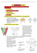
Crystal structure of KcsV channel
Left: whole structure, colour
coded with one colour for
each subunit (4 subunits).
Right: slice through the
middle showing 2 subunits,
equivalent to green and red
one, the other two subunits
having been removed to
give a better view of the
channel at the centre of the
protein.
Before this, knowledge of
channel structure and P loop was lacking. Shows that P loop forms tight
net – acts as the selectivity filter for this channel.
This selectivity filter has a
diameter of 3A, close to
sodium (2.6A).
Ion channel is an aqueous
space (water-filled cavitiy)
through a lipid membrane –
when it was crystalised, it
was in a closed
configuration (locked at the
bottom – inner helix bundle
acting as a gate).
, Potassium ions move in the opposite direction to sodium ions because
concentration is greater inside than outside the cell, so they are entering
water-filled cavity and exiting via selectivity filter.
Potassium ion (green) is surrounded by water molecules (hydration shell)
in a free state – but in the selectivity filter, there is no hydration shell.
Structure of KcsV channel reveals the mechanism of K+
selectivity
Potassium channels are specific to
potassium ions. Small diameter,
cylindrical filter that just accommodates
naked K ion.
Right: amino acid structures show red
symbols that represent oxygen atoms –
these amino acids lining selectivity filter
contribute oxygen atoms to space
which coordinate positive charged K
ions. These oxygen atoms form a cage-
like structure around the K ion,
perfectly
mimicking cage-like structure provided by
hydration shell around a free K ion.
The ‘signature sequence’ GYGVT (5 amino acids)
lines the selectivity filter with oxygen atoms (red).
These oxygens co-ordinate the K+ ion just like the
water oxygens in the ‘hydration shell’, providing a
perfect fit.
Free sodium ions are too small (1.9) for a perfect fit, but too large with
hydration shell, so are excluded from passing through
‘Signature sequence’ GYGVT
First 4 amino
acids provide an
oxygen from
their backbone
carbonyl group.
Protein structure:
2 amino acids form a peptide bond,
characterised by carbonyl and amine
group. This carbonyl oxygen is what
we see poking out into water space for
G,T,G and V.
For threonine,
hydroxyl group is its characteristic feature, which is
used to provide the oxygen required.
Structural analysis of a K+ channel to address the
questions of:
Ion selectivity
Comparison with Na+ channels
Channel gating
Voltage dependence
Inactivation
Dr Roderick MacKinnon: The Nobel Prize in Chemistry 2003
"for discoveries concerning
channels in cell membranes”
Crystal structure of a bacterial K+
channel called KcsV determined: 2
transmembrane domains and a P
loop which dips into the
membrane.
Crystal structure of KcsV channel
Left: whole structure, colour
coded with one colour for
each subunit (4 subunits).
Right: slice through the
middle showing 2 subunits,
equivalent to green and red
one, the other two subunits
having been removed to
give a better view of the
channel at the centre of the
protein.
Before this, knowledge of
channel structure and P loop was lacking. Shows that P loop forms tight
net – acts as the selectivity filter for this channel.
This selectivity filter has a
diameter of 3A, close to
sodium (2.6A).
Ion channel is an aqueous
space (water-filled cavitiy)
through a lipid membrane –
when it was crystalised, it
was in a closed
configuration (locked at the
bottom – inner helix bundle
acting as a gate).
, Potassium ions move in the opposite direction to sodium ions because
concentration is greater inside than outside the cell, so they are entering
water-filled cavity and exiting via selectivity filter.
Potassium ion (green) is surrounded by water molecules (hydration shell)
in a free state – but in the selectivity filter, there is no hydration shell.
Structure of KcsV channel reveals the mechanism of K+
selectivity
Potassium channels are specific to
potassium ions. Small diameter,
cylindrical filter that just accommodates
naked K ion.
Right: amino acid structures show red
symbols that represent oxygen atoms –
these amino acids lining selectivity filter
contribute oxygen atoms to space
which coordinate positive charged K
ions. These oxygen atoms form a cage-
like structure around the K ion,
perfectly
mimicking cage-like structure provided by
hydration shell around a free K ion.
The ‘signature sequence’ GYGVT (5 amino acids)
lines the selectivity filter with oxygen atoms (red).
These oxygens co-ordinate the K+ ion just like the
water oxygens in the ‘hydration shell’, providing a
perfect fit.
Free sodium ions are too small (1.9) for a perfect fit, but too large with
hydration shell, so are excluded from passing through
‘Signature sequence’ GYGVT
First 4 amino
acids provide an
oxygen from
their backbone
carbonyl group.
Protein structure:
2 amino acids form a peptide bond,
characterised by carbonyl and amine
group. This carbonyl oxygen is what
we see poking out into water space for
G,T,G and V.
For threonine,
hydroxyl group is its characteristic feature, which is
used to provide the oxygen required.