M.O.V.E.M.E.N.T A.L.O.N.G T.H.E. C.Y.T.O.S.K.E.L.E.T.O.N
What is a cytoskeletal motor protein?
A protein that interacts with a polarised cytoskeletal filament and moves along it using the
energy derived from ATP hydrolysis
Many types of motor proteins in eukaryotic cells which differ in the:
-> Type of filament they bind to (actin/ microtubules)
-> Direction of movement along the filament
-> The ‘cargo’ they carry (many carry organelles to their cellular locations, others cause
cytoskeletal filaments to exert tension/slide against each other to generate force for muscle
contraction, ciliary beating, cell division etc.)
Myosin II
First motor protein identified was skeletal muscle myosin, which generates the force for muscle
contraction (and is also associated, in a less organised way, with movement in various cells).
Binds cytoskeletal filaments
Elongated multi-chain protein made up of two heavy
chains (form 2 alpha helical coiled coils), each of which
associates with two different light chains (essential and
regulatory light chains)
Large globular head domain at N-terminus contains
force-generating machinery – motor head that binds to
actin filament and generates force/movement.
Followed by a very long amino acid sequence (linked to the head domain via a hinge region)
that forms an extended coiled-coil that mediates heavy chain dimerisation
Self-assembly through coiled coil tail interactions -> bundling as large bipolar filaments that
have several hundred myosin heads, orientated in opposite directions at the two ends of the
thick filament
Certain proteases can be used experimentally to cleave off the head domain – trypsin
cleaves to give a double-headed subset of the protein, and papain cleaves to isolate motor
heads from the rest of the protein.
Bipolar filament in skeletal muscle vs. non-muscle regulation
, Each myosin head binds and hydrolyses ATP, using the energy of ATP hydrolysis to walk
toward the plus end of an actin filament. The opposing orientation of the heads in the thick
filament makes the filament efficient at sliding pairs of oppositely oriented actin filaments
past eachother.
In skeletal muscle, in
which carefully
arranged actin filaments
are aligned in “thin
filament” array
surrounding the myosin
thick filaments, the
ATP-driven sliding of
actin filaments results
in muscle contraction.
Cardiac and smooth
muscle contain myosin
II molecules that are
similarly arranged,
although different
genes encode them.
Myosin II molecules aggregate by means of their tail regions, with their heads projecting to
the outside of the filament. The bare zone in the centre of the filament is free of head
domains and consists entirely of myosin II tails.
It was initially thought that myosin was present only in muscle, but in the 1970s reseaechers
found that a similar 2-headed myosin protein was also present in non-muscle cells, including
protozoan cells.
The cytoplasmic myosin II filaments in non-muscle cells are much smaller, although similarly
organised (heads on both ends and central bare zone). But importantly, these myosin II
filaments are less stable filaments – myosin is used in a more fluid/ dynamic way, with
controlled assembly when needed and then disassembly.
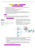
Figure: the controlled phosphorylation by the enzyme myosin light-chain kinase (MLCK) of
one of the two light chains (regulatory light chain) on non-muscle myosin II in a test tube has
at least 2 effects: it causes a change in the conformation of the myosin head, exposing its
actin-binding site, and it releases the myosin tail from a ‘sticky patch’ on the myosin head,
thereby allowing the myosin molecules to assemble into short, bipolar, thick filaments.
Motor activity contained within the myosin head region
When a muscle myosin is digested by chymotrypsin and papain, the head domain is released
as an intact fragment (called S1). The S1 fragment alone can generate filament sliding in
vitro, proving that the motor activity is contained completely within the head.
Experiment: purified S1 myosin heads
attached to a glass slide and then labelled
actin filaments were added and allowed to
bind to the myosin heads. When ATP was
added, the actin filaments began to
gradually glide along the surface in one
What is a cytoskeletal motor protein?
A protein that interacts with a polarised cytoskeletal filament and moves along it using the
energy derived from ATP hydrolysis
Many types of motor proteins in eukaryotic cells which differ in the:
-> Type of filament they bind to (actin/ microtubules)
-> Direction of movement along the filament
-> The ‘cargo’ they carry (many carry organelles to their cellular locations, others cause
cytoskeletal filaments to exert tension/slide against each other to generate force for muscle
contraction, ciliary beating, cell division etc.)
Myosin II
First motor protein identified was skeletal muscle myosin, which generates the force for muscle
contraction (and is also associated, in a less organised way, with movement in various cells).
Binds cytoskeletal filaments
Elongated multi-chain protein made up of two heavy
chains (form 2 alpha helical coiled coils), each of which
associates with two different light chains (essential and
regulatory light chains)
Large globular head domain at N-terminus contains
force-generating machinery – motor head that binds to
actin filament and generates force/movement.
Followed by a very long amino acid sequence (linked to the head domain via a hinge region)
that forms an extended coiled-coil that mediates heavy chain dimerisation
Self-assembly through coiled coil tail interactions -> bundling as large bipolar filaments that
have several hundred myosin heads, orientated in opposite directions at the two ends of the
thick filament
Certain proteases can be used experimentally to cleave off the head domain – trypsin
cleaves to give a double-headed subset of the protein, and papain cleaves to isolate motor
heads from the rest of the protein.
Bipolar filament in skeletal muscle vs. non-muscle regulation
, Each myosin head binds and hydrolyses ATP, using the energy of ATP hydrolysis to walk
toward the plus end of an actin filament. The opposing orientation of the heads in the thick
filament makes the filament efficient at sliding pairs of oppositely oriented actin filaments
past eachother.
In skeletal muscle, in
which carefully
arranged actin filaments
are aligned in “thin
filament” array
surrounding the myosin
thick filaments, the
ATP-driven sliding of
actin filaments results
in muscle contraction.
Cardiac and smooth
muscle contain myosin
II molecules that are
similarly arranged,
although different
genes encode them.
Myosin II molecules aggregate by means of their tail regions, with their heads projecting to
the outside of the filament. The bare zone in the centre of the filament is free of head
domains and consists entirely of myosin II tails.
It was initially thought that myosin was present only in muscle, but in the 1970s reseaechers
found that a similar 2-headed myosin protein was also present in non-muscle cells, including
protozoan cells.
The cytoplasmic myosin II filaments in non-muscle cells are much smaller, although similarly
organised (heads on both ends and central bare zone). But importantly, these myosin II
filaments are less stable filaments – myosin is used in a more fluid/ dynamic way, with
controlled assembly when needed and then disassembly.
Figure: the controlled phosphorylation by the enzyme myosin light-chain kinase (MLCK) of
one of the two light chains (regulatory light chain) on non-muscle myosin II in a test tube has
at least 2 effects: it causes a change in the conformation of the myosin head, exposing its
actin-binding site, and it releases the myosin tail from a ‘sticky patch’ on the myosin head,
thereby allowing the myosin molecules to assemble into short, bipolar, thick filaments.
Motor activity contained within the myosin head region
When a muscle myosin is digested by chymotrypsin and papain, the head domain is released
as an intact fragment (called S1). The S1 fragment alone can generate filament sliding in
vitro, proving that the motor activity is contained completely within the head.
Experiment: purified S1 myosin heads
attached to a glass slide and then labelled
actin filaments were added and allowed to
bind to the myosin heads. When ATP was
added, the actin filaments began to
gradually glide along the surface in one