The Eye
and the Ear
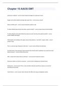
, Human eye
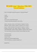
Structure of the eye
Eyeballs in bony skull sockets.
Bone, eyelids & eyelashes protect eyes from mechanical injury.
Muscles attached to eyeball keep it in socket & to move.
Glands secrete tears & oily substance onto surface prevent drying out.
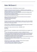
Made of three layers: sclera, choroid & retina (St Catherine’s rules)
Sclera
Outer layer, tough inelastic tissue.
Maintains shape of eyeball & is point where eye muscles attach.
Front part of sclera called cornea, transparent & allows light pass into eye.
Conjunctiva is thin membrane covers front of eyeball, contains sensory nerve endings
protect eye by detecting presence of foreign object.
Choroid
Aka middle vascular layer, dark in colour.
Contains blood vessels, brings 02 & nutrients to cells of retina. Dark colour absorbs light rays.
Prevents refraction of light inside eyeball, prevents images blurring.
Front part of choroid forms iris, coloured part.
Iris surrounds hole called pupil. Light enters eye through pupil.
Retina
Inner layer, made of photoreceptors, convert light to nerve impulses. Called rods & cones.
Rods active in dim light & allow see b&w images, contain pigment rhodopsin.
Cones active in bright light & allow see colour, pigment called iodopsin.
Fovea/yellow spot, area on retina contains only cones, area of sharp vision.
Each type of cones detects different colour, person colour blind if 1/more cones types absent.
Bipolar neurons travel from photoreceptors & leave eyeball, make blind spot.
Blood vessels enter & leave at this point. Optic nerve carries nerve impulses to visual cortex in
cerebellum where interpreted as images, allows us to see.
and the Ear
, Human eye
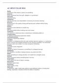
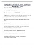
Structure of the eye
Eyeballs in bony skull sockets.
Bone, eyelids & eyelashes protect eyes from mechanical injury.
Muscles attached to eyeball keep it in socket & to move.
Glands secrete tears & oily substance onto surface prevent drying out.
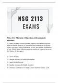
Made of three layers: sclera, choroid & retina (St Catherine’s rules)
Sclera
Outer layer, tough inelastic tissue.
Maintains shape of eyeball & is point where eye muscles attach.
Front part of sclera called cornea, transparent & allows light pass into eye.
Conjunctiva is thin membrane covers front of eyeball, contains sensory nerve endings
protect eye by detecting presence of foreign object.
Choroid
Aka middle vascular layer, dark in colour.
Contains blood vessels, brings 02 & nutrients to cells of retina. Dark colour absorbs light rays.
Prevents refraction of light inside eyeball, prevents images blurring.
Front part of choroid forms iris, coloured part.
Iris surrounds hole called pupil. Light enters eye through pupil.
Retina
Inner layer, made of photoreceptors, convert light to nerve impulses. Called rods & cones.
Rods active in dim light & allow see b&w images, contain pigment rhodopsin.
Cones active in bright light & allow see colour, pigment called iodopsin.
Fovea/yellow spot, area on retina contains only cones, area of sharp vision.
Each type of cones detects different colour, person colour blind if 1/more cones types absent.
Bipolar neurons travel from photoreceptors & leave eyeball, make blind spot.
Blood vessels enter & leave at this point. Optic nerve carries nerve impulses to visual cortex in
cerebellum where interpreted as images, allows us to see.