TASK 2
Goldstein Chapter 2:
- Short wave lengths appearing blue
- Middle wave lengths appearing green
- Long wave lengths yellow, orange and red
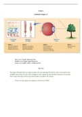
The Eye
The light reflected from an object enters the eye through the PUPIL, and is focused by the
CORNEA and LENS to form sharp images of the objects on the RETINA (network of neurons
that covers the back of the eye and contains receptors for vision)
- There are two types of receptors: RODS and CONES
,They are called ROD and CONES because their outer segments are shaped like that.
OUTER SEGMENTS contains light sensitive chemicals called VISUAL PIGMENTS that react to
light and trigger electrical signals.
These signals flow through the retina and emerge from the back of the eye in the OPTIC
NERVE.
DISTRIBITION OF RODES AND CONES:
One small area called FOVEA only contains Cones. When we look directly at an object, the
object’s image falls on the fovea.
, The Peripheral Retina (all of the retina outside of fovea) contains both Rods (12 million) and
Cones (6 million)
Macular Degeneration: destroys the cone-rich fovea and its surroundings. This creates a
blind region in central vision, when a person looks directly at something, they loses their
sight.
BLIND SPOT: The spot where nerve fibers that make up the optic nerve leaves the eye.
FOCUSING ON LIGHT:
Light reflected from an object into the eye is focused onto the retina by a two-element
optical system: THE CORNEA and THE LENS
Lens can change its shape to adjust the eye’s focus for objects located at different distances
by CILIARY MUSCLES (by increasing its curvature)
Accommodation: the change in the lens’s shape that occurs when the ciliary muscles at the
front of an eye tighten and increase the curvature of the lens so that it gets thicker. (when
looking at nearby objects)
Myopia: inability to see distant objects clearly. (the lens bends the light too much)
Goldstein Chapter 2:
- Short wave lengths appearing blue
- Middle wave lengths appearing green
- Long wave lengths yellow, orange and red
The Eye
The light reflected from an object enters the eye through the PUPIL, and is focused by the
CORNEA and LENS to form sharp images of the objects on the RETINA (network of neurons
that covers the back of the eye and contains receptors for vision)
- There are two types of receptors: RODS and CONES
,They are called ROD and CONES because their outer segments are shaped like that.
OUTER SEGMENTS contains light sensitive chemicals called VISUAL PIGMENTS that react to
light and trigger electrical signals.
These signals flow through the retina and emerge from the back of the eye in the OPTIC
NERVE.
DISTRIBITION OF RODES AND CONES:
One small area called FOVEA only contains Cones. When we look directly at an object, the
object’s image falls on the fovea.
, The Peripheral Retina (all of the retina outside of fovea) contains both Rods (12 million) and
Cones (6 million)
Macular Degeneration: destroys the cone-rich fovea and its surroundings. This creates a
blind region in central vision, when a person looks directly at something, they loses their
sight.
BLIND SPOT: The spot where nerve fibers that make up the optic nerve leaves the eye.
FOCUSING ON LIGHT:
Light reflected from an object into the eye is focused onto the retina by a two-element
optical system: THE CORNEA and THE LENS
Lens can change its shape to adjust the eye’s focus for objects located at different distances
by CILIARY MUSCLES (by increasing its curvature)
Accommodation: the change in the lens’s shape that occurs when the ciliary muscles at the
front of an eye tighten and increase the curvature of the lens so that it gets thicker. (when
looking at nearby objects)
Myopia: inability to see distant objects clearly. (the lens bends the light too much)