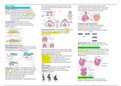
WEEK 10 REVIEW Dorsal mesocardium suspends the heart from the dorsal -no septa yet; currently 2 complete chambers, need 4
Lecture 1 - Cardiovascular System Development part (will eventually regress; still supported at the cranial -heart does beat, blood flows into the atrium from venous
Cardiogenic Mesoderm: layer of mesoderm (cardiac and caudal ends) return (umbilical vein), into ventricles, then out through
progenitor cells) induced by underlying endoderm to truncus arteriosus, dorsal aorta and to the body
become endothelial/mesodermal strands (will go on to
form heart primordium and vessels) = Angioblastic Cords;
-Positioned cranial to oropharyngeal membrane,
will need to move down; on R and L side, will
form heart primordium and fuse together to
make heart tube
-Pericardial coelom on both sides will become
pericardial cavity; some mesoderm is also forming Circulation Through Primordial Heart:
vasculature (dorsal aorta, 2 that will form along entire Initial contractions of myogenic (myocardial cells),
length of embryo) peristalsis-like waves starting at sinus venosus
Coordinated contractions begin by week 4
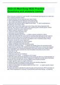
Fusion of the Heart Tube: occurs cranial to caudal, areas of Additional structures form to ensure unidirectional flow
dilation/constriction; 3 main sections formed: bulbus
cordis, ventricle, primordial atrium (need to get atria
above ventricles)
Cardiac Septation: Major septa form b/w days 27-37
Need: interatrial septum, interventricular septum, R and L
Body Folding and Heart Location: atrioventricular canals (become septum)
Cephalocaudal Folding: Rapid growth of brain vesicles pulls Partitioning of the Atrioventricular Canal:
oropharyngeal membrane forward/tucks it in; moves heart Out-pockets of tissue at ventral and dorsal border of canal
(2 tubes) towards cervical region, then thorax Fusion produces single tube with aortic and venous poles
Heart tube sprouts aortic arch vessels from aortic outflow (endocardial cushions); project into lumen and fuse, create
region, venous pole remains paired L and R AV canals that will function as the AV values;
Parts of the Heart Tube: need to fold migrate to the right of the primitive heart to serve R and L
-Bulbus Cordis - forms the right ventricle and outflow tract set of chambers
-Primitive Ventricle - forms the left ventricle
-Primitive Atrium - forms anterior parts of L and R atria
-Sinus Venosus - forms SVC and part of R atrium
-Truncus arteriosus - will help form aorta and pulmonary
trunks
Cardiac Looping:
Cephalic End (bulbus cordis) - moves ventrally, caudally,
Lateral Folding: endoderm coming together into gut tube,
and to the right beside the ventricle (d-loop)
mesoderm coming together; pericardial coeloms will come
Caudal End (atrium/sinus venosus) - moves dorsally,
together = pericardial cavity; heart tubes come together =
cranially, and to the left
tubular heart (mesoderm coming together helps bring
them together); blood going through lumen of tubular
heart (endothelial cells line the lumen)
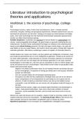
Cardiac jelly - ECM excreted by cells Interatrial Septum Formation:
Epicardium (visceral pericardium) - vs parietal pericardium Septum Primum grows from roof of common atrium;
extends to endocardial cushions; eventually fuse with it
, Opening at lower border - foramen primum; Closes up as
cell death in upper part of septum makes foramen
secundum
Septum Secundum forms in right atrium, starts to overlap
foramen secundum; form foramen ovale
Deterioration of upper part of septum leaves lower part as
the valve of the foramen ovale Lecture 2 - Vascular Development
Blood flows from RA to LA (don’t need very much blood 2 main mechanisms:
going to lungs yet - no gas exchange here); LA to LV, then -Major vessels (dorsal aorta) formed through
aorta - Some blood enters RV to pulmonary trunk but not vasculogenesis; Remainder of vascular system
much; Pressure from blood in RA opens the valve Pre/Post-Natal Foramen Ovale: around 3 months the formed through angiogenesis
valve is completely closed Pharyngeal Arches: composed of:
-Core of mesenchymal tissue derived from paraxial and
lateral mesoderm = forms muscle
-Neural crest cells migrate here = skeletal parts of face
-Surface of ectoderm and inside epithelium from
endoderm; Each arch has its own nerve and arterial supply
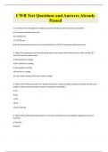
Interventricular Septum: medial walls of ventricles appose Aortic Arches: dorsal aortae have R and L branches; aortic
and merge; form muscular interventricular septum from arches branch off and connect dorsal aorta to truncus
floor of ventricle arteriosus; contribute to different arteries
Interventricular foramen on superior part - eventually -Arches appear in cranial to caudal sequence; NOT all
closed d/t outgrowth of endocardial cushions downward arches contribute to vessels
and bulbus cordis (bulbar ridges) -Separation through the aortic sac (ventral aorta) will
occur in cranial to caudal direction
Development of Bulbus Cordis: forms outflow tracts of
both ventricles; truncus arteriosus contributes to great
vessels
-Mesenchymal cells in bulbus cordis form bulbar ridges; Development of the Vascular Pattern:
form roots of aorta and pulmonary artery
-Neural crest cells from truncal ridges in truncus arteriosus
(migrate through pharyngeal arches)
-Opposing ridges grow towards each and spiral 180
degrees, separate the outflow tracts of the RV and LV =
form aorticopulmonary septum - Blood from RV needs to
go to pulmonary trunk; blood from LV needs to go to aortic
trunk
Lecture 1 - Cardiovascular System Development part (will eventually regress; still supported at the cranial -heart does beat, blood flows into the atrium from venous
Cardiogenic Mesoderm: layer of mesoderm (cardiac and caudal ends) return (umbilical vein), into ventricles, then out through
progenitor cells) induced by underlying endoderm to truncus arteriosus, dorsal aorta and to the body
become endothelial/mesodermal strands (will go on to
form heart primordium and vessels) = Angioblastic Cords;
-Positioned cranial to oropharyngeal membrane,
will need to move down; on R and L side, will
form heart primordium and fuse together to
make heart tube
-Pericardial coelom on both sides will become
pericardial cavity; some mesoderm is also forming Circulation Through Primordial Heart:
vasculature (dorsal aorta, 2 that will form along entire Initial contractions of myogenic (myocardial cells),
length of embryo) peristalsis-like waves starting at sinus venosus
Coordinated contractions begin by week 4
Fusion of the Heart Tube: occurs cranial to caudal, areas of Additional structures form to ensure unidirectional flow
dilation/constriction; 3 main sections formed: bulbus
cordis, ventricle, primordial atrium (need to get atria
above ventricles)
Cardiac Septation: Major septa form b/w days 27-37
Need: interatrial septum, interventricular septum, R and L
Body Folding and Heart Location: atrioventricular canals (become septum)
Cephalocaudal Folding: Rapid growth of brain vesicles pulls Partitioning of the Atrioventricular Canal:
oropharyngeal membrane forward/tucks it in; moves heart Out-pockets of tissue at ventral and dorsal border of canal
(2 tubes) towards cervical region, then thorax Fusion produces single tube with aortic and venous poles
Heart tube sprouts aortic arch vessels from aortic outflow (endocardial cushions); project into lumen and fuse, create
region, venous pole remains paired L and R AV canals that will function as the AV values;
Parts of the Heart Tube: need to fold migrate to the right of the primitive heart to serve R and L
-Bulbus Cordis - forms the right ventricle and outflow tract set of chambers
-Primitive Ventricle - forms the left ventricle
-Primitive Atrium - forms anterior parts of L and R atria
-Sinus Venosus - forms SVC and part of R atrium
-Truncus arteriosus - will help form aorta and pulmonary
trunks
Cardiac Looping:
Cephalic End (bulbus cordis) - moves ventrally, caudally,
Lateral Folding: endoderm coming together into gut tube,
and to the right beside the ventricle (d-loop)
mesoderm coming together; pericardial coeloms will come
Caudal End (atrium/sinus venosus) - moves dorsally,
together = pericardial cavity; heart tubes come together =
cranially, and to the left
tubular heart (mesoderm coming together helps bring
them together); blood going through lumen of tubular
heart (endothelial cells line the lumen)
Cardiac jelly - ECM excreted by cells Interatrial Septum Formation:
Epicardium (visceral pericardium) - vs parietal pericardium Septum Primum grows from roof of common atrium;
extends to endocardial cushions; eventually fuse with it
, Opening at lower border - foramen primum; Closes up as
cell death in upper part of septum makes foramen
secundum
Septum Secundum forms in right atrium, starts to overlap
foramen secundum; form foramen ovale
Deterioration of upper part of septum leaves lower part as
the valve of the foramen ovale Lecture 2 - Vascular Development
Blood flows from RA to LA (don’t need very much blood 2 main mechanisms:
going to lungs yet - no gas exchange here); LA to LV, then -Major vessels (dorsal aorta) formed through
aorta - Some blood enters RV to pulmonary trunk but not vasculogenesis; Remainder of vascular system
much; Pressure from blood in RA opens the valve Pre/Post-Natal Foramen Ovale: around 3 months the formed through angiogenesis
valve is completely closed Pharyngeal Arches: composed of:
-Core of mesenchymal tissue derived from paraxial and
lateral mesoderm = forms muscle
-Neural crest cells migrate here = skeletal parts of face
-Surface of ectoderm and inside epithelium from
endoderm; Each arch has its own nerve and arterial supply
Interventricular Septum: medial walls of ventricles appose Aortic Arches: dorsal aortae have R and L branches; aortic
and merge; form muscular interventricular septum from arches branch off and connect dorsal aorta to truncus
floor of ventricle arteriosus; contribute to different arteries
Interventricular foramen on superior part - eventually -Arches appear in cranial to caudal sequence; NOT all
closed d/t outgrowth of endocardial cushions downward arches contribute to vessels
and bulbus cordis (bulbar ridges) -Separation through the aortic sac (ventral aorta) will
occur in cranial to caudal direction
Development of Bulbus Cordis: forms outflow tracts of
both ventricles; truncus arteriosus contributes to great
vessels
-Mesenchymal cells in bulbus cordis form bulbar ridges; Development of the Vascular Pattern:
form roots of aorta and pulmonary artery
-Neural crest cells from truncal ridges in truncus arteriosus
(migrate through pharyngeal arches)
-Opposing ridges grow towards each and spiral 180
degrees, separate the outflow tracts of the RV and LV =
form aorticopulmonary septum - Blood from RV needs to
go to pulmonary trunk; blood from LV needs to go to aortic
trunk