MODULE 10 GENERAL HISTOLOGICAL ORGANIZATION OF THE DIGESTIVE
TRACT WALL
HOW ARE YOU DIGESTING ANAT
Although individual parts of the digestive tract perform
100? THE DIGESTIVE SYSTEM distinct functions, there are structural similarities when
comparing the histology.
SECTION 01: COMPONENTS OF THE DIGESTIVE Learn about the histological layers of the digestive tract
SYSTEM using the esophagus as an example.
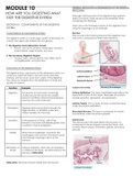
COMPONENTS OF THE DIGESTIVE SYSTEM Mucosa
The mucosa is the innermost layer of the digestive tract
The digestive system is a multi-organ system of the body. As wall, and is composed of three main elements.
a result, the organs are divided into two groups:
1. The Digestive Tract (Alimentary Canal):
• Mouth, oral cavity, pharynx, esophagus, stomach,
small intestine, large intestine, and anus
2. The Accessory Digestive Organs:
• Tongue, teeth, salivary glands, liver, biliary ducts
and gallbladder, and pancreas
FUNCTIONS OF THE DIGESTIVE TRACT
The digestive tract, also known as the alimentary canal, is a
continuous tube that starts in the oral cavity and ends
inferiorly with the anal canal. Each organ of the digestive
tract plays an integral role in the breakdown of food and
uptake of nutrients.
Function Example
Digestion The process of mastication (chewing) Surface Epithelium: The type of epithelial layer reflects the
crushes food in the mouth into smaller expected function of the organ. Examples of functions
pieces to facilitate chemical processing by include secretion, absorption, and protection.
enzymes into small molecules.
Lamina Propria: This is a layer of loose connective tissue
under the surface epithelium.
Absorption Digested food moves slowly through the
large intestine, to facilitate water and
Muscularis Mucosa: This layer is composed of smooth
nutrient uptake into the body.
muscle fibres under the lamina propria.
Secretion In the presence of food, cells of the Submucosa
stomach’s mucosal wall release gastric acid The submucosa is the layer below the mucosa. It is
to perform chemical digestion. composed of dense irregular connective tissue, and
contains blood vessels, lymphatics, glands, and nerve
plexuses.
Motility When food is swallowed, muscles in the
esophageal wall contract and relax to push
food through esophagus down to the
stomach.
Elimination of Leftover materials, which are not absorbed
Waste or utilized by the body, are eliminated by the
process of defecation.
Defecation: Removal of fecal matter from the rectum.
, Muscularis Externa SECTION 02: ORAL CAVITY
The muscularis externa is composed of circular and
longitudinal layers of smooth muscle with nerve plexuses in
INTRODUCTION TO THE ORAL CAVITY
between the layers.
The oral cavity is the first part of the digestive tract. It
The plexuses are responsible for muscular contraction to
consists of two parts:
propel food through the digestive tract.
• The vestibule, which is the space between the
cheeks and the lips and the gums and teeth
• The oral cavity proper, which includes the other
areas of the mouth
Serosa/ Adventitia
The outermost layer of the digestive tract is either a serosa
or an adventitia.
When the outer layer is a serous membrane, it is known as a ORAL CAVITY
serosa. When the outer layer is composed of loose
connective tissue, it is known as adventitia. 1. PALATE
The palate makes up the superior border of the oral cavity.
Serous membrane: A single layer of thin, flat cells that form It is divided into the hard palate (bone) and soft palate
a membranous sheet and secrete lubricating fluid. (muscle). The posterior extension of the soft palate is called
the uvula.
2. TONGUE
The tongue is a muscle associated with speech, taste, and
the mechanical manipulation of food.
REVIEW: LAYERS OF THE DIGESTIVE TRACT WALL
The tongue is made up of muscles that control the shape of
Layer Definition the tongue itself (intrinsic muscles), and muscles that move
the tongue during chewing and speech (extrinsic muscles).
Mucosa Composed of surface epithelium,
lamina propria, and muscularis On the inferior surface of the tongue is the frenulum, which
mucosa. anchors the tongue to the floor of the mouth.
Submucosa A layer of dense irregular connective
tissue with blood vessels, nerves,
lymphatics, glands, etc.
Muscularis Externa Circular and longitudinal layers of
smooth muscle that account for
peristalsis and segmentation.
Serosa or
Adventitia Layer that is either serous in nature, or
composed of loose connective tissue.
TRACT WALL
HOW ARE YOU DIGESTING ANAT
Although individual parts of the digestive tract perform
100? THE DIGESTIVE SYSTEM distinct functions, there are structural similarities when
comparing the histology.
SECTION 01: COMPONENTS OF THE DIGESTIVE Learn about the histological layers of the digestive tract
SYSTEM using the esophagus as an example.
COMPONENTS OF THE DIGESTIVE SYSTEM Mucosa
The mucosa is the innermost layer of the digestive tract
The digestive system is a multi-organ system of the body. As wall, and is composed of three main elements.
a result, the organs are divided into two groups:
1. The Digestive Tract (Alimentary Canal):
• Mouth, oral cavity, pharynx, esophagus, stomach,
small intestine, large intestine, and anus
2. The Accessory Digestive Organs:
• Tongue, teeth, salivary glands, liver, biliary ducts
and gallbladder, and pancreas
FUNCTIONS OF THE DIGESTIVE TRACT
The digestive tract, also known as the alimentary canal, is a
continuous tube that starts in the oral cavity and ends
inferiorly with the anal canal. Each organ of the digestive
tract plays an integral role in the breakdown of food and
uptake of nutrients.
Function Example
Digestion The process of mastication (chewing) Surface Epithelium: The type of epithelial layer reflects the
crushes food in the mouth into smaller expected function of the organ. Examples of functions
pieces to facilitate chemical processing by include secretion, absorption, and protection.
enzymes into small molecules.
Lamina Propria: This is a layer of loose connective tissue
under the surface epithelium.
Absorption Digested food moves slowly through the
large intestine, to facilitate water and
Muscularis Mucosa: This layer is composed of smooth
nutrient uptake into the body.
muscle fibres under the lamina propria.
Secretion In the presence of food, cells of the Submucosa
stomach’s mucosal wall release gastric acid The submucosa is the layer below the mucosa. It is
to perform chemical digestion. composed of dense irregular connective tissue, and
contains blood vessels, lymphatics, glands, and nerve
plexuses.
Motility When food is swallowed, muscles in the
esophageal wall contract and relax to push
food through esophagus down to the
stomach.
Elimination of Leftover materials, which are not absorbed
Waste or utilized by the body, are eliminated by the
process of defecation.
Defecation: Removal of fecal matter from the rectum.
, Muscularis Externa SECTION 02: ORAL CAVITY
The muscularis externa is composed of circular and
longitudinal layers of smooth muscle with nerve plexuses in
INTRODUCTION TO THE ORAL CAVITY
between the layers.
The oral cavity is the first part of the digestive tract. It
The plexuses are responsible for muscular contraction to
consists of two parts:
propel food through the digestive tract.
• The vestibule, which is the space between the
cheeks and the lips and the gums and teeth
• The oral cavity proper, which includes the other
areas of the mouth
Serosa/ Adventitia
The outermost layer of the digestive tract is either a serosa
or an adventitia.
When the outer layer is a serous membrane, it is known as a ORAL CAVITY
serosa. When the outer layer is composed of loose
connective tissue, it is known as adventitia. 1. PALATE
The palate makes up the superior border of the oral cavity.
Serous membrane: A single layer of thin, flat cells that form It is divided into the hard palate (bone) and soft palate
a membranous sheet and secrete lubricating fluid. (muscle). The posterior extension of the soft palate is called
the uvula.
2. TONGUE
The tongue is a muscle associated with speech, taste, and
the mechanical manipulation of food.
REVIEW: LAYERS OF THE DIGESTIVE TRACT WALL
The tongue is made up of muscles that control the shape of
Layer Definition the tongue itself (intrinsic muscles), and muscles that move
the tongue during chewing and speech (extrinsic muscles).
Mucosa Composed of surface epithelium,
lamina propria, and muscularis On the inferior surface of the tongue is the frenulum, which
mucosa. anchors the tongue to the floor of the mouth.
Submucosa A layer of dense irregular connective
tissue with blood vessels, nerves,
lymphatics, glands, etc.
Muscularis Externa Circular and longitudinal layers of
smooth muscle that account for
peristalsis and segmentation.
Serosa or
Adventitia Layer that is either serous in nature, or
composed of loose connective tissue.