Group 1 (Water contact angle)
- Quantative measurement (determine wettability of a surface by water).
- Indication of hydrophilicity (smaller angle indicates hydrophilic surface)
- Wetting/ non-wetting (complete not wetting (180 degrees) complete wetting (90)
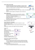
- Static contact angles (boundary is not moving)
- Dynamic contact angles (no stationary contact angles, 2 angles can be distinguished
(picture)
- Factors to consider (roughness, heterogeneity, purity of solvent etc.)
Group 2 (Ellipsometry)
- Measures thickness and optical constant of thin films, with high
precision. It measures the change of polarization upon reflection or
transmission in comparison to a model.
- Characterize composition, roughness, thickness etc.
- Different fields use this
- Advantages: measures at least two parameters of each wavelength & measures an
intensity ratio instead of intensities.
Elliptical polarization of light is used.
Group 3 (Atomic force microscopy)
Sharp tip attached to a cantilever(silicon) which is attached to a z-piezo
(nanometer range accuracy)
Bending of cantilever (by repulsive energy) changes the direction of the laser
beam
Feedback loop: variation surface height and results in photodiode
Contact AFM:
Simple
Strong repulsive forces
Deformation or damage to surface
Easy to interpret
Non-Contact AFM:
AKA. Dynamic force microscopy
5-15 nm above surface
No deformation
Amplitude to determine surface
Time delay in feedback loop (disadvantage)
Group 4 (Laser scanning confocal microscopy)
Capabilities of CLSM
- Focus on a single focal plane within the specimen achieving sharp images
- From these single planes a 3D image can be obtained (stack sliced images)
- -> Provides structural and organizational information about cells and tissues
Advantages of CLSM
Compared to widefield fluorescence microscopy
- Control over the depth of field (because uses specific wavelengths)
, - Reduction of information that is not in the focal plane
- Allows to slice thicker specimens
Compared to TEM
- 3D imaging (TEM doesn’t allow this)
- Easier specimen preparation
Compared to SEM
- No need for vacuum condition (with CLSM)
- More detail, not only surface (CLSM can give more than only surface)
Compared to AFM
- No risk of tip interfering with specimen for CLSM
Group 5 (Dynamic light scattering)
- Technique in physics used to determine the size of (macromolecules)
particles down to 1 nm in diameter. The basic principle of DLS: 1. The sample
is illuminated by a laser beam and the fluctuations of the scattered light are
detected at a known scattering angle (θ) by a fast photon detector.
Basic principles
- Brownian motion: particles are constantly colliding with solvent molecules collisions
cause a certain amount of energy to be transferred – induces particle movement
- Smaller particles are moving and diffusing at higher speed than larger particles
- The relation between the speed of the particles and the particle size is given by the
stokes-Einstein equation – the speed of the particles is given by the translational
diffusion coefficient D – Requirement: particles move solely based on Brownian
motion -> no sedimentation
- Size limits: 1. Upper size: sedimentation 2. Lower size: signal-to-noise ratio
Advantages and disadvantages
Advantages:
● non-invasive, fast, and automated
● modest development costs
● can analyze samples containing broad distribution of species with different molecular
masses
● can detect small amounts of higher mass species (<0.01% in many cases)
suitable for molecular weight determination and size measurements of molecules in the
range of 10µm to less than 1 nm
● can also obtain radius of gyration and the translational diffusion coefficient
Disadvantages and Limitations:
● highly sensitive to solvent viscosity and temperature
● low resolution method ⇒ cannot differentiate between closely related molecules (e.g.
monomer and dimer)
● can only analyze liquid dispersion, whereas laser diffraction is effective for dry powders
too
● presence of large aggregates significantly affect the measurements
- Quantative measurement (determine wettability of a surface by water).
- Indication of hydrophilicity (smaller angle indicates hydrophilic surface)
- Wetting/ non-wetting (complete not wetting (180 degrees) complete wetting (90)
- Static contact angles (boundary is not moving)
- Dynamic contact angles (no stationary contact angles, 2 angles can be distinguished
(picture)
- Factors to consider (roughness, heterogeneity, purity of solvent etc.)
Group 2 (Ellipsometry)
- Measures thickness and optical constant of thin films, with high
precision. It measures the change of polarization upon reflection or
transmission in comparison to a model.
- Characterize composition, roughness, thickness etc.
- Different fields use this
- Advantages: measures at least two parameters of each wavelength & measures an
intensity ratio instead of intensities.
Elliptical polarization of light is used.
Group 3 (Atomic force microscopy)
Sharp tip attached to a cantilever(silicon) which is attached to a z-piezo
(nanometer range accuracy)
Bending of cantilever (by repulsive energy) changes the direction of the laser
beam
Feedback loop: variation surface height and results in photodiode
Contact AFM:
Simple
Strong repulsive forces
Deformation or damage to surface
Easy to interpret
Non-Contact AFM:
AKA. Dynamic force microscopy
5-15 nm above surface
No deformation
Amplitude to determine surface
Time delay in feedback loop (disadvantage)
Group 4 (Laser scanning confocal microscopy)
Capabilities of CLSM
- Focus on a single focal plane within the specimen achieving sharp images
- From these single planes a 3D image can be obtained (stack sliced images)
- -> Provides structural and organizational information about cells and tissues
Advantages of CLSM
Compared to widefield fluorescence microscopy
- Control over the depth of field (because uses specific wavelengths)
, - Reduction of information that is not in the focal plane
- Allows to slice thicker specimens
Compared to TEM
- 3D imaging (TEM doesn’t allow this)
- Easier specimen preparation
Compared to SEM
- No need for vacuum condition (with CLSM)
- More detail, not only surface (CLSM can give more than only surface)
Compared to AFM
- No risk of tip interfering with specimen for CLSM
Group 5 (Dynamic light scattering)
- Technique in physics used to determine the size of (macromolecules)
particles down to 1 nm in diameter. The basic principle of DLS: 1. The sample
is illuminated by a laser beam and the fluctuations of the scattered light are
detected at a known scattering angle (θ) by a fast photon detector.
Basic principles
- Brownian motion: particles are constantly colliding with solvent molecules collisions
cause a certain amount of energy to be transferred – induces particle movement
- Smaller particles are moving and diffusing at higher speed than larger particles
- The relation between the speed of the particles and the particle size is given by the
stokes-Einstein equation – the speed of the particles is given by the translational
diffusion coefficient D – Requirement: particles move solely based on Brownian
motion -> no sedimentation
- Size limits: 1. Upper size: sedimentation 2. Lower size: signal-to-noise ratio
Advantages and disadvantages
Advantages:
● non-invasive, fast, and automated
● modest development costs
● can analyze samples containing broad distribution of species with different molecular
masses
● can detect small amounts of higher mass species (<0.01% in many cases)
suitable for molecular weight determination and size measurements of molecules in the
range of 10µm to less than 1 nm
● can also obtain radius of gyration and the translational diffusion coefficient
Disadvantages and Limitations:
● highly sensitive to solvent viscosity and temperature
● low resolution method ⇒ cannot differentiate between closely related molecules (e.g.
monomer and dimer)
● can only analyze liquid dispersion, whereas laser diffraction is effective for dry powders
too
● presence of large aggregates significantly affect the measurements