Primary Muscular Disorders
•More than 600 separate muscles
•40% of the human weight in adulthood
•A muscle contains thousands of muscle fibers that extend for variable distances along
its longitudinal axis.
•A muscular fiber is a multinucleated cell from a few mm to several cm and from 10-100
micrometers diameter
•Muscle fibers innervated by an anterior horn cell act as a unit called –“motor unit” –
basic physiologic unit in all reflex, postural and voluntar activity.
•A particular muscle may have from a few (3-4 as extraocular muscles) to hundreds of
MU (like the cvadriceps muscle)
Clinical syndrome
•Motor deficit not restricted to a particular motor unit.
•Distribution of motor weakness might involve a certain group of muscles or could be
more widespread. In this case proximal muscles are usually more involved –wadling gait
is characteristic for pelvian muscles deficit.
•Distal myopaties are not quite exceptional.
•Muscle tone is diminished
•Muscular atrophy and loss of bulck is common
•Common activities might get impaired because of weakness related to a
muscular disorder:
–Walking, running, climbing stairs, arising from sitting (special sign –Gowers –“patient
climbing on himself”), kneeling, squatting or reclining position, working with hands above
shoulder’s level.
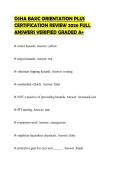
--Localized muscle weakness might be a sign of a muscular disorder as well: drooping of
the eyelids, diplopia and strabismus, change in facial expression and voice, difficulty in
chewing, closing the mouth, swallowing etc..
1
,•Non muscular abnormalities in muscular diseases
–Dislocation of the hips
–Skin involvement
–Cardiac abnormalities
–Retinian changes
–Cerebellum and cranial nerves
–Fronto-temporal dementia
–Dysmorphic features of cranium
–Endocrine abnormalities
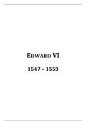
Skin lesions –typical rash in dermatomyositis
Differential diagnosis with polineuropathies
•Tendon reflexes are spared
•No sensory signs
•No sphincterian troubles
•No autonomic signs
But we can have:
pain, spasms, cramps, rippling, muscular hypertrophy (true or false), fatigue, myotonia,
stiffness, muscle mass or change in muscle volume
Diagnosis
•Blood tests
•Electrophysiology
•Imaging studies
•Biopsy
2
,•Genetic tests
•Asses cardiac function for associated cardiomiopathy
Blood tests
•Check up for:
–Creatinkinase (elevated in active, progressive disorders to thousands of units)
–Aldolase
–Transaminases
–Electrolites: sodium, potasium, cloride
–Tiroidian check up
–Autoimmune battery
–Toxicology
–Serology for viral or parasitic infections
Electrophysiology
•Nerve conduction studies are normal
•Needle electrode test of muscular activity at rest and gradual to maximal contraction
–At rest we can find:
fibrillation potentials -markers for spontaneous contractions of individual muscle
fibers as a consequence of membrane instability–very prominent in inflammatory
myopathies during the active episodes
myotonic discharge -repetitive high frequency potentials expression of delayed
muscle relaxation after voluntary contraction–action myotonia or mechanical
stimulation–percussion myotonia Types Chloride channel-related disorders–eg, myotonia
congenita, Thomsen type; protein kinase-related disorders–eg, myotonic dystrophy;
sodium channel-related disorders–eg, hyperkalemic periodic paralysis; idiopathic
3
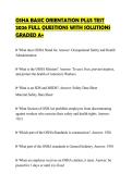
, •Normal recruitment
•Early recruitment during gradual contraction in myopathic conditions with low
amplitude and short duration of individual motor units
•Myopathic pattern of amplitude/turn ratio (under the lower part of the cloud)
4
•More than 600 separate muscles
•40% of the human weight in adulthood
•A muscle contains thousands of muscle fibers that extend for variable distances along
its longitudinal axis.
•A muscular fiber is a multinucleated cell from a few mm to several cm and from 10-100
micrometers diameter
•Muscle fibers innervated by an anterior horn cell act as a unit called –“motor unit” –
basic physiologic unit in all reflex, postural and voluntar activity.
•A particular muscle may have from a few (3-4 as extraocular muscles) to hundreds of
MU (like the cvadriceps muscle)
Clinical syndrome
•Motor deficit not restricted to a particular motor unit.
•Distribution of motor weakness might involve a certain group of muscles or could be
more widespread. In this case proximal muscles are usually more involved –wadling gait
is characteristic for pelvian muscles deficit.
•Distal myopaties are not quite exceptional.
•Muscle tone is diminished
•Muscular atrophy and loss of bulck is common
•Common activities might get impaired because of weakness related to a
muscular disorder:
–Walking, running, climbing stairs, arising from sitting (special sign –Gowers –“patient
climbing on himself”), kneeling, squatting or reclining position, working with hands above
shoulder’s level.
--Localized muscle weakness might be a sign of a muscular disorder as well: drooping of
the eyelids, diplopia and strabismus, change in facial expression and voice, difficulty in
chewing, closing the mouth, swallowing etc..
1
,•Non muscular abnormalities in muscular diseases
–Dislocation of the hips
–Skin involvement
–Cardiac abnormalities
–Retinian changes
–Cerebellum and cranial nerves
–Fronto-temporal dementia
–Dysmorphic features of cranium
–Endocrine abnormalities
Skin lesions –typical rash in dermatomyositis
Differential diagnosis with polineuropathies
•Tendon reflexes are spared
•No sensory signs
•No sphincterian troubles
•No autonomic signs
But we can have:
pain, spasms, cramps, rippling, muscular hypertrophy (true or false), fatigue, myotonia,
stiffness, muscle mass or change in muscle volume
Diagnosis
•Blood tests
•Electrophysiology
•Imaging studies
•Biopsy
2
,•Genetic tests
•Asses cardiac function for associated cardiomiopathy
Blood tests
•Check up for:
–Creatinkinase (elevated in active, progressive disorders to thousands of units)
–Aldolase
–Transaminases
–Electrolites: sodium, potasium, cloride
–Tiroidian check up
–Autoimmune battery
–Toxicology
–Serology for viral or parasitic infections
Electrophysiology
•Nerve conduction studies are normal
•Needle electrode test of muscular activity at rest and gradual to maximal contraction
–At rest we can find:
fibrillation potentials -markers for spontaneous contractions of individual muscle
fibers as a consequence of membrane instability–very prominent in inflammatory
myopathies during the active episodes
myotonic discharge -repetitive high frequency potentials expression of delayed
muscle relaxation after voluntary contraction–action myotonia or mechanical
stimulation–percussion myotonia Types Chloride channel-related disorders–eg, myotonia
congenita, Thomsen type; protein kinase-related disorders–eg, myotonic dystrophy;
sodium channel-related disorders–eg, hyperkalemic periodic paralysis; idiopathic
3
, •Normal recruitment
•Early recruitment during gradual contraction in myopathic conditions with low
amplitude and short duration of individual motor units
•Myopathic pattern of amplitude/turn ratio (under the lower part of the cloud)
4